Journal of Traditional Chinese Medicine ›› 2022, Vol. 42 ›› Issue (6): 900-907.DOI: 10.19852/j.cnki.jtcm.20220815.001
• Research Articles • Previous Articles Next Articles
Efficacy of Dangua Fang (丹瓜方) on endothelial cells damaged by oxidative stress
HENG Xianpei( ), LI Liang, YANG Liuqin, WANG Zhita
- Department of Endocrinology, People's Hospital Affiliated to Fujian University of Traditional Chinese Medicine, Fuzhou 350004, China
-
Received:2021-07-07Accepted:2021-10-17Online:2022-12-15Published:2022-08-15 -
Contact:HENG Xianpei,LI Liang,YANG Liuqin -
About author:Prof. HENG Xianpei, Department of Endocrinology, People's Hospital Affiliated to Fujian University of Traditional Chinese Medicine, Fuzhou 350004, China. hengxianpei@hotmail.com, Telephone: +86-13067366157
-
Supported by:Based on The "miR34a/Nampt-NAD+-TAC" Pathway to Study the Mechanism of Simultaneously Treating The Phlegm And Blood Stasis in The Regulation of Glycolipid(81873213);Study on the Mechanism of Simultaneously Treating the Phlegm and Blood Stasis on Glycolipid Metabolism Based on Intestinal Fat Absorption Regulated by miR-34a/Stat3-Nfil3 Pathway(82074308);Preparation of Monomeric Traditional Chinese Medicine Complexes Based on Nampt's Activation of Tricarboxylic Acid Cycle And Respiratory Chain to Interfere with Glycolipid Metabolism(2022Y41016)
Cite this article
HENG Xianpei, LI Liang, YANG Liuqin, WANG Zhita. Efficacy of Dangua Fang (丹瓜方) on endothelial cells damaged by oxidative stress[J]. Journal of Traditional Chinese Medicine, 2022, 42(6): 900-907.
share this article
Table 1 Comparison of OD values of different concentrations of Dangua Fang-containing serum intervention of endothelial cell proliferation activity injure induced by oxidative stress ($\bar{x} \pm s$)
| Group | n | OD value | ReR (%) | Group | n | OD value | ReR (%) | Group | n | OD value | ReR (%) |
|---|---|---|---|---|---|---|---|---|---|---|---|
| Control | 3 | 0.5772±0.0321 | - | DG3 | 3 | 0.4113±0.0536c | 32.268 | DD1 | 3 | 0.3224±0.0113 | –7.076 |
| Model | 3 | 0.3429±0.0313a | - | DZ1 | 3 | 0.4356±0.0213c | 42.393 | DD2 | 3 | 0.3542±0.0024 | 5.934 |
| DG1 | 3 | 0.3778±0.0144 | 19.392 | DZ2 | 3 | 0.4242±0.0334c | 34.104 | DD3 | 3 | 0.3755±0.0157 | 19.323 |
| DG2 | 3 | 0.3851±0.0329b | 20.478 | DZ3 | 3 | 0.3266±0.0278 | –1.977 |
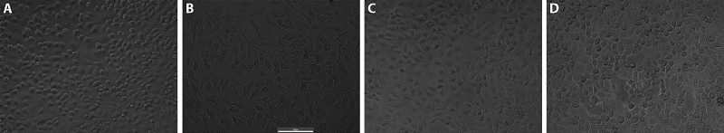
Figure 1 oxidative stress-induced injured endothelial cells under different interventions (×200) Figures were photographed under an inverted microscope. A: conventional culture, no intervention; B: without drug intervention after modelling (1000μmol/L final concentration of H2O2-caused oxidative stress-damage); C: after modelling, treatment with Dangua Fang drug-containing serum; D: medium containing final concentrations of vitamin C of 20 μg/mL and coenzyme Q of 80 μg/mL. cells were cultured for 48 h.
Figure 1 oxidative stress-induced injured endothelial cells under different interventions (×200) Figures were photographed under an inverted microscope. A: conventional culture, no intervention; B: without drug intervention after modelling (1000μmol/L final concentration of H2O2-caused oxidative stress-damage); C: after modelling, treatment with Dangua Fang drug-containing serum; D: medium containing final concentrations of vitamin C of 20 μg/mL and coenzyme Q of 80 μg/mL. cells were cultured for 48 h.
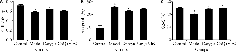
Figure 2 Comparison of cell viability, apoptosis and cell cycle of oxidative stress-induced injured cell (n = 6,$\bar{x} \pm s$) A: comparison of cell viability; B: comparison of apoptosis; C: comparison of G2 + S. Control: conventional culture, no intervention; Model: without drug intervention after modelling (1000μmol/L final concentration of H2O2-caused oxidative stress-damage); Dangua: after modelling, treatment with Dangua Fang drug-containing serum; CoQ + Vit C: medium containing final concentrations of vitamin C of 20 μg/mL and coenzyme Q of 80 μg/mL. Cells were cultured for 48 h. compared with Control group: aP < 0.05; compared with Model group: bP < 0.01, cP < 0.05. G2 + S: Late stage of DNA synthesis + DNA synthesis stage.
Figure 2 Comparison of cell viability, apoptosis and cell cycle of oxidative stress-induced injured cell (n = 6,$\bar{x} \pm s$) A: comparison of cell viability; B: comparison of apoptosis; C: comparison of G2 + S. Control: conventional culture, no intervention; Model: without drug intervention after modelling (1000μmol/L final concentration of H2O2-caused oxidative stress-damage); Dangua: after modelling, treatment with Dangua Fang drug-containing serum; CoQ + Vit C: medium containing final concentrations of vitamin C of 20 μg/mL and coenzyme Q of 80 μg/mL. Cells were cultured for 48 h. compared with Control group: aP < 0.05; compared with Model group: bP < 0.01, cP < 0.05. G2 + S: Late stage of DNA synthesis + DNA synthesis stage.
Table 2 Influence of Dangua Fang on mitochondrial respiratory chain enzymes (μg/μL, $\bar{x} \pm s$)
| Group | n | SDH | COX |
|---|---|---|---|
| Control | 3 | 9.73±1.13 | 1.11±0.07 |
| Model | 3 | 11.91±0.77a | 0.78±0.04a |
| Dangua | 3 | 12.63±0.30 | 0.90±0.07c |
| CoQ+VitC | 3 | 6.92±0.18b | 0.82±0.04 |
Table 3 Comparison of Mitochondrial Membrane Potential Parameters in all groups ($\bar{x} \pm s$)
| Group | n | R3 Red/green (×100) | R2 Red/green (×100) | R1 Red/green (×100) | Green (R2 + R3) |
|---|---|---|---|---|---|
| Contol | 4 | 13.502±0.579 | 58.104±0.843 | 45.366±3.911 | 695.294±11.408 |
| Model | 4 | 3.658±0.012a | 47.802±0.502a | 6.513±0.130a | 887.108±3.724a |
| Dangua | 4 | 6.836±0.014b | 60.835±0.199b | 51.015±4.365b | 638.714±25.206b |
| CoQ+VitC | 4 | 10.129±0.009b | 73.697±1.041b | 61.643±0.636b | 686.668±14.191b |
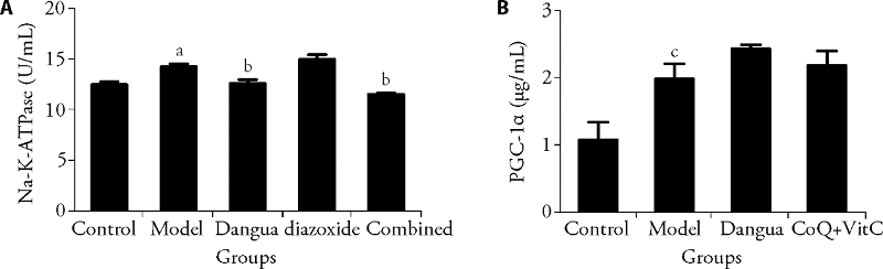
Figure 3 Comparison of Na+-k+-ATPase and PGC-1αin all groups (n = 3, $\bar{x} \pm s$) A: comparison of Na+-k+-ATPase: Control group: conventional culture, no intervention; Model group: without drug intervention after modelling (1000μmol/L final concentration of H2O2-caused oxidative stress-damage); Dangua group: after modelling, treatment with Dangua Fang drug-containing serum; Diazoxide group: the culture solution contained a final diazoxide concentration of 0.1 μmol/mL; Combined: diazoxide + Dangua Fang. B: Comparsion of PGC-1α. The treatments of the control group, model group and Dangua group were the same as in Figure 3A. CoQ + Vit C group: medium containing final concentrations of vitamin C of 20 μg/mL and coenzyme Q of 80 μg/mL. Cells were cultured for 48 h. Compared with Control group, aP < 0.01, cP < 0.05; compared with Model group, bP < 0.01. PGC-1α: peroxisome proliferators-activated receptor-γcoactivator-1.
Figure 3 Comparison of Na+-k+-ATPase and PGC-1αin all groups (n = 3, $\bar{x} \pm s$) A: comparison of Na+-k+-ATPase: Control group: conventional culture, no intervention; Model group: without drug intervention after modelling (1000μmol/L final concentration of H2O2-caused oxidative stress-damage); Dangua group: after modelling, treatment with Dangua Fang drug-containing serum; Diazoxide group: the culture solution contained a final diazoxide concentration of 0.1 μmol/mL; Combined: diazoxide + Dangua Fang. B: Comparsion of PGC-1α. The treatments of the control group, model group and Dangua group were the same as in Figure 3A. CoQ + Vit C group: medium containing final concentrations of vitamin C of 20 μg/mL and coenzyme Q of 80 μg/mL. Cells were cultured for 48 h. Compared with Control group, aP < 0.01, cP < 0.05; compared with Model group, bP < 0.01. PGC-1α: peroxisome proliferators-activated receptor-γcoactivator-1.
| [1] | Heng XP, Yang LQ, Li L, Chen ML. Paradox of using intensive lowering of blood glucose in diabetics and strategies to overcome it and decrease cardiovascular risks. Chin J Integr Med 2015, 21: 425-34 |
| [2] |
Heng XP, Li XJ, Li L, Yang LQ, Wang ZT. Therapy to obese Type 2 diabetes mellitus: how far will we go down the wrong road? Chin J Integr Med 2020, 26: 62-71.
DOI URL |
| [3] | Pistritto G, Trisciuoglio D, Ceci C, Garufi A, D'Orazi G. Apoptosis as anticancer mechanism: function and dysfunction of its modulators and targeted therapeutic strategies. Aging (Albany NY) 2016; 8: 603-19. |
| [4] |
Chen L, Yang W, Guo Y, et al. Exosomal lncRNA GAS 5 regulates the apoptosis of macrophages and vascular endothelial cells in atherosclerosis. PLoS One 2017; 12: e0185406.
DOI URL |
| [5] |
Watson EC, Grant ZL, Coultas L. Endothelial cell apoptosis in angiogenesis and vessel regression. Cell Mol Life Sci 2017; 74: 4387-403.
DOI PMID |
| [6] |
Lan YL, Huang XP, Heng XP, et al. Dangua Fang improves glycolipid metabolic disorders by promoting hepatic adenosine 5’-monophosphate activated protein kinase expression in diabetic Goto-Kakizaki rats. Chin J Integr Med 2015; 21: 188-95.
DOI URL |
| [7] | Huang SP, Kang WQ, Liu YJ, Huang BW, Shao JJ. Effects of Dangua formula on the protein expression of LKB1, AMPK and SIRT1 in liver of diabetic rats. Zhong Hua Zhong Yi Yao Za Zhi 2019; 34: 4003-7. |
| [8] | Heng XP, Huang SP, Cheng XL, et al. Research of Dangua recipe on intervening the glycolipid metabolism and oxidative stress in diabetic rats with atherosclerosis. Zhong Guo Zhong Xi Yi Jie He Za Zhi 2013; 33: 244-51. |
| [9] | Heng XP, Li L, Huang SP, et al. Effect of Dangua recipe on glycolipid metabolism and VCAM-1 and its mRNA expression level in Apo E (-/-) mice with diabetes mellitus. Zhong Guo Zhong Xi Yi Jie He Za Zhi 2014; 34: 1086-95. |
| [10] | Yang LQ, Li L, Heng XP, Huang SP, Pan XD. Effects of Dangua recipe on inflammeatory marders and endothelial cell function in diabetic rats with arteriosclerosis. Zhong Guo Zhong Xi Yi Jie He Za Zhi 2017, 37: 692-8. |
| [11] | Xu RX, Wang ZT, Cheng YC, et al. Effects of Dangua recipe on myocardial ATP, PPARα, GLUT-4,and morphology in diabetic rats. Zhong Guo Zhong Xi Yi Jie He Za Zhi 2018; 38: 1363-8. |
| [12] | Chen YC, Li L, Heng XP, et al. Effects of Dangua recipe on expression leveis of caspase-3 protein, Bcl-2 and Bax mRNA in brain tissue of Apo E-/- diabetes model mice. Zhong Guo Zhong Xi Yi Jie He Za Zhi 2017; 37: 1476-81. |
| [13] | Heng XP, Yang LQ, Li L, Pan XD, Huang SP. Dangua recipe regulates protein and mRNA expression of p38-MAPK, MCP-1 and FN in renal of Apo E-/-murine model of diabetes. Zhong Guo Zhong Xi Yi Jie He Za Zhi 2018; 39: 459-65. |
| [14] | Heng XP, Yang LQ, Huang SP, et al. A Clinical study of Danggua Humai oral liquid on cardiovascular risk factors among patients with type 2 diabetes mellitus. Zhong Guo Zhong Xi Yi Jie He Za Zhi 2019; 39: 275-81. |
| [15] |
Heng XP, Chen KJ, Hong ZF, et al. Glucose endothelial cytotoxicity and protection of Dan Gua-Fang, a Chinese herb prescription in huVEC in hyperglycemia medium. J Diabetes Complications 2009, 23: 297-303.
DOI URL |
| [16] |
Heng XP, Chen KJ, Hong ZF, et al. Toxicity features of high glucose on endothelial cell cycle and protection by Dan Gua-Fang in ECV-304 in high glucose medium. Chin J Integr Med. 2013, 19: 596-602.
DOI URL |
| [17] |
Gu MX, Wang J, Wang Y, et al. MiR-147b inhibits cell viability and promotes apoptosis of rat H9c2 cardiomyocytes via down-regulating KLF13 expression. Acta Biochim Biophys Sin (Shanghai) 2018; 50: 288-97.
DOI URL |
| [18] | Liao GL, Ma L, Zeng T. The effects medlar flavone on no and NOS in H2O2damaged vascular endothelial cell. Chong Qin Yi Xue 2015, 44: 3323-27 |
| [19] | Tian G, Liu ZQ, Yuan ZY. Membrance phospholipids injury in myocardia cells of adriamycin rats and the protective effects of CoQ10 on it. Xi An Yi Ke Da Xue Xue Bao 1997, 18: 329-33 |
| [20] | Cropper JR, Hicks M, Ryan JB, Macdonald PS. Enhanced cardioprotection of the rat heart during hypothermic storage with combined Na+ - H+ exchange inhibition and ATP-dependent potassium channel activation. J Heart Lung Transplant World J Tradit Chin Med 2003; 22: 1245-53. |
| [21] |
Wada J, Nakatsuka A. Mitochondrial dynamics and mitochondrial dysfunction in diabetes. Acta Med Okayama 2016; 70: 151-8.
PMID |
| [22] |
Rocha M, Diaz-Morales N, Rovira-Llopis S, et al. Mitochondrial dysfunction and endoplasmic reticulum stress in diabetes. Curr Pharm Des 2016; 22: 2640-9.
DOI URL |
| [23] |
Gu XL.MicroRNA-124 prevents H2O2-induced apoptosis and oxidative stress in human lens epithelial cells via inhibition of the NF-κB signaling pathway. Pharmacology 2018; 102: 213-22.
DOI URL |
| [24] |
Holvoet P, Vanhaverbeke M, Geeraert B, De Keyzer D, Hulsmans M, Janssens S. Low cytochrome oxidase 1 links mitochondrial dysfu nction to atherosclerosis in mice and pigs. PLoS One 2017; 12: e0170307.
DOI URL |
| [25] |
Sarparanta J, García-Macia M, Singh R.Autophagy and mitochondria in obesity and type 2 diabetes. Curr Diabetes Rev 2017; 13: 352-9.
DOI PMID |
| [26] |
Kogot-Levin A, Saada A, Leibowitz G, et al. Upregulation of mitochondrial content in cytochrome c oxidase deficient fibroblasts. PLoS One 2016; 11: e0165417.
DOI URL |
| [27] |
De La Fuente S, Lambert JP, Nichtova Z, et al. Spatial separation of mitochondrial calcium uptake and extrusion for energy-efficient mitochondrial calcium signaling in the heart. Cell Rep 2018; 24: 3099- 107. e4.
DOI PMID |
| [28] |
Li PA, Hou X, Hao S. Mitochondrial biogenesis in neurodegeneration. J Neurosci Res 2017; 95: 2025-9.
DOI PMID |
| [29] |
Heng XP, Wang ZT, Li L, Yang LQ, Huang SP. Mechanisms of Dangua recipe in improving glycolipid metabolic disorders based on transcriptomics. Chin J Integr Med 2022; 28: 130-7.
DOI URL |
| [30] | Cantó C, Auwerx J. PGC-1alpha, SIRT1 and AMPK, an energy sensing network that controls energy expenditure. Curr Opin Lipidol 2009; 20: 98-105. |
| [31] |
Tang BL. Sirt1 and the mitochondria. Mol Cells 2016; 39: 87-95.
DOI PMID |
| [32] |
Shaw RJ, Lamia KA, Vasquez D, et al. The kinase LKB1 mediates glucose homeostasis in liver and therapeutic effects of metformin. Science 2005; 310: 1642-6.
DOI PMID |
| [1] | HUANG Hongmei, YANG Maojun, LI Ting, WANG Dandan, LI Ying, TANG Xiaochi, YUAN Lu, GU Shi, XU Yong. Neferine inhibits the progression of diabetic nephropathy by modulating the miR-17-5p/nuclear factor E2-related factor 2 axis [J]. Journal of Traditional Chinese Medicine, 2024, 44(1): 44-53. |
| [2] | HENG Xianpei, WANG Zhita, YANG Liuqing, LI Liang, HUANG Suping. Dangua Fang (丹瓜方) regulating tricarboxylic acid cycle and respiratory chain and its mechanism in diabetic rats [J]. Journal of Traditional Chinese Medicine, 2023, 43(6): 1150-1159. |
| [3] | ZHANG Xiaoying, WANG Ruixuan, WANG Yiqing, XU Fanxing, YAN Tingxu, WU Bo, ZHANG Ming, JIA Ying. Spinosin protects Neuro-2a/APP695 cells from oxidative stress damage by inactivating p38 [J]. Journal of Traditional Chinese Medicine, 2023, 43(5): 868-875. |
| [4] | LIU Bingbing, LI Jieru, SI Jianchao, CHEN Qi, YANG Shengchang, JI Ensheng. Ginsenoside Rb1 alleviates chronic intermittent hypoxia-induced diabetic cardiomyopathy in db/db mice by regulating the adenosine monophosphate-activated protein kinase/Nrf2/heme oxygenase-1 signaling pathway [J]. Journal of Traditional Chinese Medicine, 2023, 43(5): 906-914. |
| [5] | ZHOU Hua, LI Hui, WANG Haihua. Potential protective effects of the water-soluble Chinese propolis on experimental ulcerative colitis [J]. Journal of Traditional Chinese Medicine, 2023, 43(5): 925-933. |
| [6] | WU Haiyang, WANG Ying, HAN Wei, LI Huihui, JI Haisheng, LIU Xiuxiu. Protective effect of Tongdu Tiaoshen acupuncture combined with Xiaoxuming decoction (小续命汤) on dopaminergic neurons in Parkinson’s disease model [J]. Journal of Traditional Chinese Medicine, 2023, 43(3): 484-493. |
| [7] | JIANG Wen, ZHANG Wei, ZHANG Yuxiang, YANG Hao, PAN Xiaomei, CHEN Qiang, CHEN Junhui. Tilianin extracted from Xiangqinglan (Herba Dracocephali Moldovicae) inhibits apoptosis induced by mitochondrial pathway and endoplasmic reticulum stress in H9c2 cells after oxygen-glucose deprivation/reoxygenation [J]. Journal of Traditional Chinese Medicine, 2023, 43(1): 42-50. |
| [8] | LI Han, HUANG Xiaomin, CAI Haiyang, HEROK George, HE Jing, SU Yixun, LI Weihong, YI Chenju, OLIVER Brian G, CHEN Hui. Mitochondrial dysfunction in a rat model and the related risk of metabolic disorders [J]. Journal of Traditional Chinese Medicine, 2023, 43(1): 95-104. |
| [9] | ZHENG Wei, WANG Mingxing, LIU Shanxue, LUAN Chao, ZHANG Yanqiu, XU Duoduo, WANG Jian. Buyang Huanwu Tang (补阳还五汤) protects H2O2-induced RGC-5 cell against oxidative stress and apoptosis via reactive oxygen species-mitogen-activated protein kinase signaling pathway [J]. Journal of Traditional Chinese Medicine, 2022, 42(6): 885-891. |
| [10] | ZHU Lingyan, WEI Yihong, WANG Youhua, YANG Jianmei, LI Jiawei, CAO Min, ZHOU Duan. Protective efficacy of Shenge San (参蛤散) on mitochondria in H9c2 cardiomyocytes [J]. Journal of Traditional Chinese Medicine, 2022, 42(6): 892-899. |
| [11] | HUANG Qiuyue, YE Hui, SHI Zongming, JIA Xiaofen, LIN Miaomiao, CHU Yingming, YU Jing, ZHANG Xuezhi. Efficacy of Qingre Huashi decoction (清热化湿方) on infection of Helicobacter pylori: inhibiting adhesion, antioxidant, and anti-inflammation [J]. Journal of Traditional Chinese Medicine, 2022, 42(6): 915-921. |
| [12] | CHEN Jinpeng, ZHANG Kexia, LIU Yi, JIN Song, GAI Xiaohong, REN Tao, TIAN Chengwang. Efficacy of phospholipid complex of flavonoids from persimmon leaves on atherosclerosis, and possible mechanism [J]. Journal of Traditional Chinese Medicine, 2022, 42(3): 417-425. |
| [13] | ZHAO Lixia, SUN Wei, BAI Decheng. Protective effect of resveratrol on rat cardiomyocyte H9C2 cells injured by hypoxia/reoxygenation by regulating mitochondrial autophagy via PTEN-induced putative kinase protein 1/Parkinson disease protein 2 signaling pathway [J]. Journal of Traditional Chinese Medicine, 2022, 42(2): 176-186. |
| [14] | Xing DU, Tianlong LIU, Wendi TAO, Maoxing LI, Xiaolin LI, Lan YAN. Effect of aqueous extract of Astragalus membranaceus on behavioral cognition of rats living at high altitude [J]. Journal of Traditional Chinese Medicine, 2022, 42(1): 58-64. |
| [15] | ZHU Guohua, SUN Xipeng, DING Cuntao, ZHAO Huan, LI Jing, HUA Qi. Effect of Songlingxuemaikang(松龄血脉康) on mild essential hypertension in patients: a randomized parallel-controlled study [J]. Journal of Traditional Chinese Medicine, 2021, 41(5): 799-805. |
| Viewed | ||||||
|
Full text |
|
|||||
|
Abstract |
|
|||||