
Journal of Traditional Chinese Medicine ›› 2026, Vol. 46 ›› Issue (5): 1206-1217.DOI: 10.19852/j.cnki.jtcm.2026.05.014
• Original Articles • Previous Articles Next Articles
Jiangtang Xiaozhi formula (降糖消脂方) alleviates type 2 diabetes mellitus by improving islet beta-cell dedifferentiation: a network pharmacology-based study
ZHU Meilin1, DU Jianpeng2, LUO Yunpeng3, LIU Yunlu4, NIU Zenghui4, LAN Tian1, YANG Jing4, CHEN Zhuhong1( )
- 1
Department of Endocrinology ,Xiyuan Hospital, China Academy of Chinese Medical Sciences Beijing 100091, China
2Cardiovascular Diseases Center ,Xiyuan Hospital, China Academy of Chinese Medical Sciences Beijing 100091, China
3Department of Endocrinology ,Dongzhimen Hospital, Beijing University of Chinese Medicine Beijing 100700, China
4Beijing Key Laboratory of Traditional Chinese Medicine Basic Research on Prevention and Treatment for Major Diseases ,Experimental Research Center, China Academy of Chinese Medical Sciences Beijing 100700, China
-
Received:2025-04-04Accepted:2026-01-27Online:2026-10-15Published:2026-10-09 -
Contact:CHEN Zhuhong, Department of Endocrinology, Xiyuan Hospital, China Academy of Chinese Medical Sciences, Beijing 100091, China. 13264214919@163.com,Telephone: +86-17852938921 -
Supported by:Science and Technology Innovation Project of China Academy of Chinese Medical Sciences: Study on the Mechanism of Jiangtang Xiaozhi Formula in Treating Diabetes by Regulating Pancreatic Beta-cell Dedifferentiation via the Forkhead Box O1-Mediated Insulin-like Growth Factor 1/Phosphatidylinositol 3-Kinase/Protein Kinase B Pathway(Grant Number: CI2021A01618);Special Project for Research And Development And Transformation of Intellectual Property Medical Institution Preparations and New Chinese Medicines of Xiyuan Hospital, China Academy of Chinese Medical Sciences: a Randomized, Double-Blind, Placebo-Controlled Clinical Study of Jiangtang Xiaozhi Granules for the Treatment of Metabolic Dysfunction-Associated Fatty Liver Disease(Grant Number: XYZX0303-01);Youth Quest Project of China Association of Chinese Medicine: a Clinical Study of Jiangtang Xiaozhi Formula for the Treatment of Type 2 Diabetes Mellitus Complicated with Non-Alcoholic Fatty Liver Disease(Grant Number: 2023-QNQS-07);National Natural Science Foundation of China: Molecular Mechanism of Yiqi Huoxue Formula in Delaying Vascular Inflammatory Aging Based on the Angiotensin II/Angiotensin II Type 1 Receptor/Nuclear Factor Kappa B Inflammatory Pathway(Grant Number: 82074260)

Cite this article
ZHU Meilin, DU Jianpeng, LUO Yunpeng, LIU Yunlu, NIU Zenghui, LAN Tian, YANG Jing, CHEN Zhuhong. Jiangtang Xiaozhi formula (降糖消脂方) alleviates type 2 diabetes mellitus by improving islet beta-cell dedifferentiation: a network pharmacology-based study[J]. Journal of Traditional Chinese Medicine, 2026, 46(5): 1206-1217.
share this article
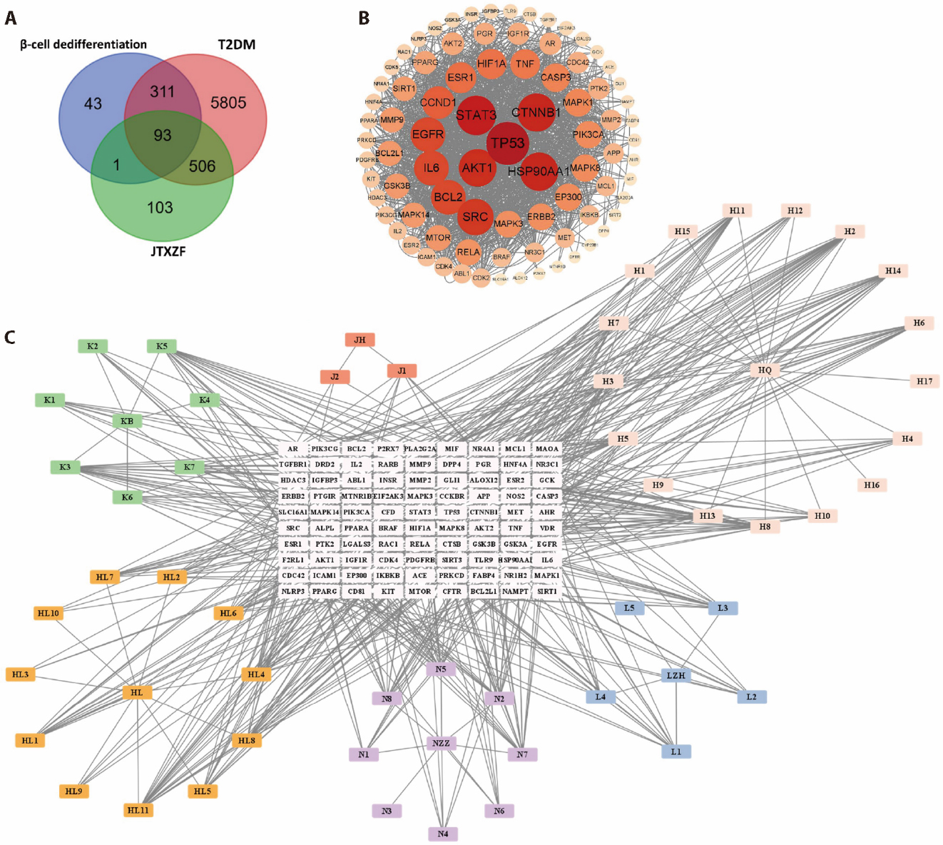
Figure 1 Network pharmacology analysis A: venn diagram of overlapping targets of T2DM, Islet beta-cell dedifferentiation and JTXZF; B: PPI network of 93 overlapping genes of T2DM, Islet beta-cell dedifferentiation and JTXZF; C: regarding the drug-component-target network of JTXZF in relation to T2DM and beta-cell dedifferentiation. T2DM: type 2 diabetes mellitus; PPI: protein-protein interaction; JTXZF: Jiangtang Xiaozhi formula.
Figure 1 Network pharmacology analysis A: venn diagram of overlapping targets of T2DM, Islet beta-cell dedifferentiation and JTXZF; B: PPI network of 93 overlapping genes of T2DM, Islet beta-cell dedifferentiation and JTXZF; C: regarding the drug-component-target network of JTXZF in relation to T2DM and beta-cell dedifferentiation. T2DM: type 2 diabetes mellitus; PPI: protein-protein interaction; JTXZF: Jiangtang Xiaozhi formula.
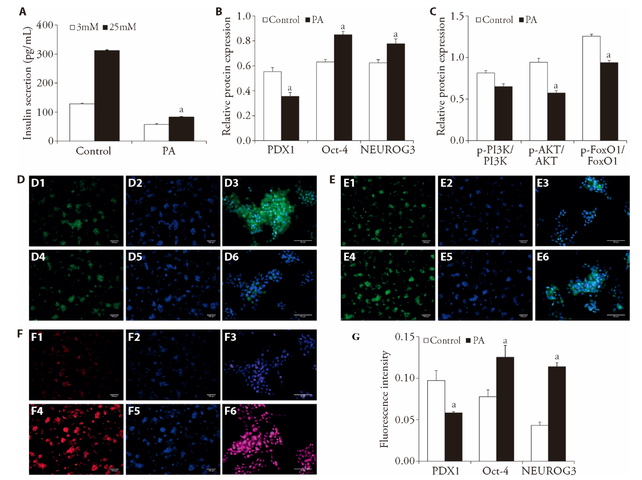
Figure 2 Inhibition of the PI3K/AKT/FoxO1 signaling pathway mediates PA-induced islet beta-cell dedifferentiation A: Min6 cells were cultured in 3 and 25 mM glucose before measuring insulin concentrations of the supernatant using a commercial ELISA kit; B: densitometric quantification of Western blot bands for PDX1, Oct-4 and NEUROG3, normalized to β-Actin; C: densitometric quantification of Western blot bands for p-PI3K/PI3K, p-AKT/AKT and p-FoxO1/FoxO1, normalized to β-Actin; D: PDX1 expression in Min6 cells was detected by immunofluorescence staining; D1, D2, D4, D5: original magnification, × 200; scale bar = 100 μm; D3, D6: original magnification, × 400; scale bar = 50 μm; E: Oct-4 expression in Min6 cells was detected by immunofluorescence staining; E1, E2, E4, E5: original magnification, × 200; scale bar = 100 μm; E3, E6: original magnification, × 400; scale bar = 50 μm; F: NEURGO3 expression in Min6 cells was detected by immunofluorescence staining; F1, F2, F4, F5: original magnification, × 200; scale bar = 100 μm; F3, F6: original magnification, × 400; scale bar = 50 μm; G: fluorescence intensity for PDX1, Oct-4 and NEUROG3; D1, D2, D3, E1, E2, E3, F1, F2, F3: Control group; D4, D5, D6, E4, E5, E6, F4, F5, F6: PA group. Control group: normal group; PA group: cells were treated with 0.3 mM palmitic acid for 24 h. PA: palmitic acid; PDX1: pancreatic and duodenal homeobox 1; Oct-4: octamer-binding transcription factor 4; NEUROG3: neurogenin 3; p-PI3K/PI3K: phosphorylated phosphoinositide 3-kinase/total phosphoinositide 3-kinase; p-AKT/AKT: phosphorylated protein kinase B/total protein kinase B; p-FoxO1/FoxO1: phosphorylated forkhead box O1/total forkhead box O1. Statistical comparisons between two groups were conducted using unpaired t-tests, while multiple group comparisons were performed using one-way analysis of variance, followed by Tukey’s test for pairwise multiple comparisons. All data are expressed as mean ± standard error of the mean (n = 3). aP < 0.05 vs respective Control.
Figure 2 Inhibition of the PI3K/AKT/FoxO1 signaling pathway mediates PA-induced islet beta-cell dedifferentiation A: Min6 cells were cultured in 3 and 25 mM glucose before measuring insulin concentrations of the supernatant using a commercial ELISA kit; B: densitometric quantification of Western blot bands for PDX1, Oct-4 and NEUROG3, normalized to β-Actin; C: densitometric quantification of Western blot bands for p-PI3K/PI3K, p-AKT/AKT and p-FoxO1/FoxO1, normalized to β-Actin; D: PDX1 expression in Min6 cells was detected by immunofluorescence staining; D1, D2, D4, D5: original magnification, × 200; scale bar = 100 μm; D3, D6: original magnification, × 400; scale bar = 50 μm; E: Oct-4 expression in Min6 cells was detected by immunofluorescence staining; E1, E2, E4, E5: original magnification, × 200; scale bar = 100 μm; E3, E6: original magnification, × 400; scale bar = 50 μm; F: NEURGO3 expression in Min6 cells was detected by immunofluorescence staining; F1, F2, F4, F5: original magnification, × 200; scale bar = 100 μm; F3, F6: original magnification, × 400; scale bar = 50 μm; G: fluorescence intensity for PDX1, Oct-4 and NEUROG3; D1, D2, D3, E1, E2, E3, F1, F2, F3: Control group; D4, D5, D6, E4, E5, E6, F4, F5, F6: PA group. Control group: normal group; PA group: cells were treated with 0.3 mM palmitic acid for 24 h. PA: palmitic acid; PDX1: pancreatic and duodenal homeobox 1; Oct-4: octamer-binding transcription factor 4; NEUROG3: neurogenin 3; p-PI3K/PI3K: phosphorylated phosphoinositide 3-kinase/total phosphoinositide 3-kinase; p-AKT/AKT: phosphorylated protein kinase B/total protein kinase B; p-FoxO1/FoxO1: phosphorylated forkhead box O1/total forkhead box O1. Statistical comparisons between two groups were conducted using unpaired t-tests, while multiple group comparisons were performed using one-way analysis of variance, followed by Tukey’s test for pairwise multiple comparisons. All data are expressed as mean ± standard error of the mean (n = 3). aP < 0.05 vs respective Control.
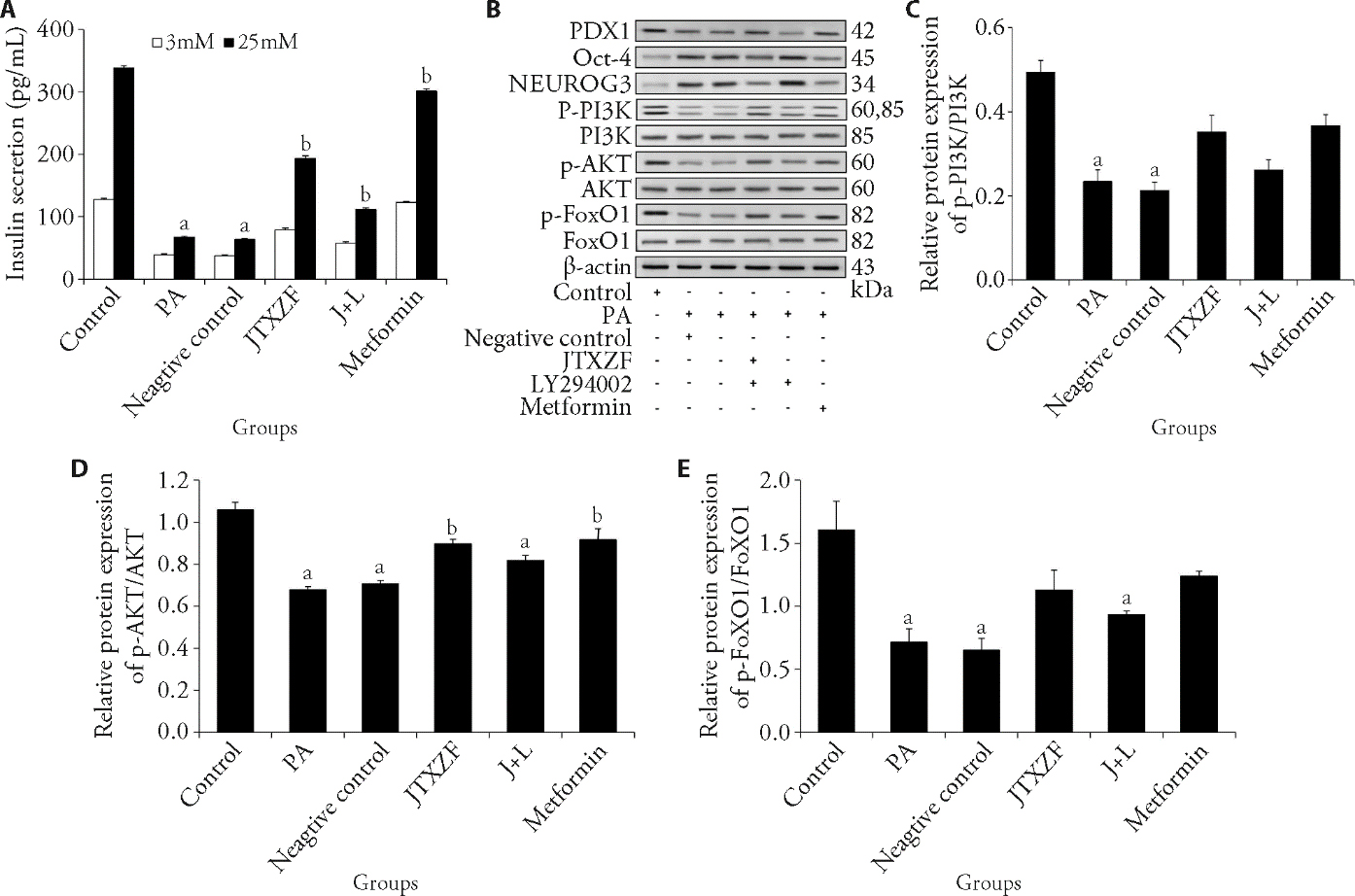
Figure 3 JTXZF alleviates PA-induced dysfunction and dedifferentiation in Min6 cells via the PI3K/Akt/FoxO1 signaling pathway A: Min6 cells were cultured in 3 and 25 mM glucose before measuring insulin concentrations of the supernatant using a commercial ELISA kit; B: protein electrophoresis of PDX1, Oct-4, NEUROG3 and PI3K/AKT/FoxO1 signaling molecule; C: densitometric quantification of Western blot bands for p-PI3K/PI3K, normalized to β-Actin; D: densitometric quantification of Western blot bands for p-AKT/AKT, normalized to β-actin. E: densitometric quantification of Western blot bands for p-FoxO1/FoxO1, normalized to β-Actin. control group: normal group; PA group: cells were treated with 0.3 mM palmitic acid for 24 h; negative control group: PA-treated cells received 20% blank serum for 24 h; JTXZF group: PA-treated cells received 20% JTXZF-containing serum for 24 h; J + L group: Cells were treated with 0.3 mM PA, 20% Jiangtang Xiaozhi formula-containing serum, and 5 μM PI3K inhibitor LY294002 for 24 h; Metformin group: Cells were treated with 0.3 mM PA and 200 μM metformin. PA: palmitic acid; JTXZF: Jiangtang Xiaozhi formula; J + L: JTXZF + PI3K inhibitor LY294002; PDX1: pancreatic and duodenal homeobox 1; Oct-4: octamer-binding transcription factor 4; NEUROG3: neurogenin 3; p-PI3K/PI3K: phosphorylated phosphoinositide 3-kinase/total phosphoinositide 3-kinase; p-AKT/AKT: phosphorylated protein kinase B/total protein kinase B; p-FoxO1/FoxO1: phosphorylated forkhead box O1/total forkhead box O1.Statistical comparisons between two groups were conducted using unpaired t-tests, while multiple group comparisons were performed using one-way analysis of variance, followed by Tukey’s test for pairwise multiple comparisons. All data are expressed as mean ± standard error of the mean (n = 3). aP < 0.05 vs respective Control; bP < 0.05 vs respective PA.
Figure 3 JTXZF alleviates PA-induced dysfunction and dedifferentiation in Min6 cells via the PI3K/Akt/FoxO1 signaling pathway A: Min6 cells were cultured in 3 and 25 mM glucose before measuring insulin concentrations of the supernatant using a commercial ELISA kit; B: protein electrophoresis of PDX1, Oct-4, NEUROG3 and PI3K/AKT/FoxO1 signaling molecule; C: densitometric quantification of Western blot bands for p-PI3K/PI3K, normalized to β-Actin; D: densitometric quantification of Western blot bands for p-AKT/AKT, normalized to β-actin. E: densitometric quantification of Western blot bands for p-FoxO1/FoxO1, normalized to β-Actin. control group: normal group; PA group: cells were treated with 0.3 mM palmitic acid for 24 h; negative control group: PA-treated cells received 20% blank serum for 24 h; JTXZF group: PA-treated cells received 20% JTXZF-containing serum for 24 h; J + L group: Cells were treated with 0.3 mM PA, 20% Jiangtang Xiaozhi formula-containing serum, and 5 μM PI3K inhibitor LY294002 for 24 h; Metformin group: Cells were treated with 0.3 mM PA and 200 μM metformin. PA: palmitic acid; JTXZF: Jiangtang Xiaozhi formula; J + L: JTXZF + PI3K inhibitor LY294002; PDX1: pancreatic and duodenal homeobox 1; Oct-4: octamer-binding transcription factor 4; NEUROG3: neurogenin 3; p-PI3K/PI3K: phosphorylated phosphoinositide 3-kinase/total phosphoinositide 3-kinase; p-AKT/AKT: phosphorylated protein kinase B/total protein kinase B; p-FoxO1/FoxO1: phosphorylated forkhead box O1/total forkhead box O1.Statistical comparisons between two groups were conducted using unpaired t-tests, while multiple group comparisons were performed using one-way analysis of variance, followed by Tukey’s test for pairwise multiple comparisons. All data are expressed as mean ± standard error of the mean (n = 3). aP < 0.05 vs respective Control; bP < 0.05 vs respective PA.

Figure 4 JTXZF upregulates IGF-1 to activate the PI3K/Akt/FoxO1 signaling pathway, thereby alleviating PA-induced dysfunction and dedifferentiation in MIN6 cells A: the Min6 cells expression of IGF-1 mRNA quantified by RT-qPCR; B: Min6 cells were cultured in 3 and 25mM glucose before measuring insulin concentrations of the supernatant using a commercial ELISA kit; C: fluorescence intensity for IGF-1; D: IGF-1 expression in Min6 cells was detected by immunofluorescence staining; D1, D2, D4, D5, D7, D8, D10, D11: original magnification, ×200; scale bar = 100 μm; D3, D6, D9, D12: original magnification, × 400; scale bar = 50 μm; E: protein electrophoresis of IGF-1, and PI3K/AKT/FoxO1 signaling molecule; F: densitometric quantification of Western blot bands for IGF-1, normalized to β-Actin; G: densitometric quantification of Western blot bands for p-PI3K/PI3K, p-AKT/AKT and p-FoxO1/FoxO1, normalized to β-Actin; D1, D2, D3: Control group; D4, D5, D6: PA group; D7, D8, D9: JTXZF group; D10, D11, D12: J + si-IGF-1 group. Control group: normal group; PA group: cells were treated with 0.3 mM palmitic acid for 24 h; JTXZF group: PA-treated cells received 20% Jiangtang Xiaozhi formula -containing serum for 24 h; J + si-IGF-1 group: Cells were treated with 0.3 mM PA and 20% JTXZF-containing serum and transfected with small interfering RNA targeting IGF-1 for 24 h. PA: palmitic acid; JTXZF: Jiangtang Xiaozhi formula; J + si-IGF-1: JTXZF + IGF-1 inhibitor group; IGF-1: insulin-like growth factor 1; p-PI3K/PI3K: phos-phorylated phosphoinositide 3-kinase/total phosphoinositide 3-kinase; p-AKT/AKT: phosphorylated protein kinase B/total protein kinase B; p-FoxO1/FoxO1: phosphorylated forkhead box O1/total forkhead box O1; RT-qPCR: real-time quantitative polymerase chain reaction. Statistical comparisons between two groups were conducted using unpaired t-tests, while multiple group comparisons were performed using one-way analysis of variance, followed by Tukey’s test for pairwise multiple comparisons. All data are expressed as mean ± standard error of the mean (n = 3). aP < 0.05 vs respective Control; bP < 0.05 vs respective PA.
Figure 4 JTXZF upregulates IGF-1 to activate the PI3K/Akt/FoxO1 signaling pathway, thereby alleviating PA-induced dysfunction and dedifferentiation in MIN6 cells A: the Min6 cells expression of IGF-1 mRNA quantified by RT-qPCR; B: Min6 cells were cultured in 3 and 25mM glucose before measuring insulin concentrations of the supernatant using a commercial ELISA kit; C: fluorescence intensity for IGF-1; D: IGF-1 expression in Min6 cells was detected by immunofluorescence staining; D1, D2, D4, D5, D7, D8, D10, D11: original magnification, ×200; scale bar = 100 μm; D3, D6, D9, D12: original magnification, × 400; scale bar = 50 μm; E: protein electrophoresis of IGF-1, and PI3K/AKT/FoxO1 signaling molecule; F: densitometric quantification of Western blot bands for IGF-1, normalized to β-Actin; G: densitometric quantification of Western blot bands for p-PI3K/PI3K, p-AKT/AKT and p-FoxO1/FoxO1, normalized to β-Actin; D1, D2, D3: Control group; D4, D5, D6: PA group; D7, D8, D9: JTXZF group; D10, D11, D12: J + si-IGF-1 group. Control group: normal group; PA group: cells were treated with 0.3 mM palmitic acid for 24 h; JTXZF group: PA-treated cells received 20% Jiangtang Xiaozhi formula -containing serum for 24 h; J + si-IGF-1 group: Cells were treated with 0.3 mM PA and 20% JTXZF-containing serum and transfected with small interfering RNA targeting IGF-1 for 24 h. PA: palmitic acid; JTXZF: Jiangtang Xiaozhi formula; J + si-IGF-1: JTXZF + IGF-1 inhibitor group; IGF-1: insulin-like growth factor 1; p-PI3K/PI3K: phos-phorylated phosphoinositide 3-kinase/total phosphoinositide 3-kinase; p-AKT/AKT: phosphorylated protein kinase B/total protein kinase B; p-FoxO1/FoxO1: phosphorylated forkhead box O1/total forkhead box O1; RT-qPCR: real-time quantitative polymerase chain reaction. Statistical comparisons between two groups were conducted using unpaired t-tests, while multiple group comparisons were performed using one-way analysis of variance, followed by Tukey’s test for pairwise multiple comparisons. All data are expressed as mean ± standard error of the mean (n = 3). aP < 0.05 vs respective Control; bP < 0.05 vs respective PA.
| [1] | Cole JB, Florez JC. Genetics of diabetes mellitus and diabetes complications. Nat Rev Nephrol 2020; 16: 377-90. |
| [2] | Ahmad E, Lim S, Lamptey R, Webb DR, Davies MJ. Type 2 diabetes. Lancet 2022; 400: 1803-20. |
| [3] | Eizirik DL, Pasquali L, Cnop M. Pancreatic β-cells in type 1 and type 2 diabetes mellitus: different pathways to failure. Nat Rev Endocrinol 2020; 16: 349-62. |
| [4] | Hudish LI, Reusch JE, Sussel L. beta cell dysfunction during progression of metabolic syndrome to type 2 diabetes. J Clin Invest 2019; 129: 4001-8. |
| [5] | Ghorbani A, Rashidi R, Shafiee-Nick R. Flavonoids for preserving pancreatic beta cell survival and function: a mechanistic review. Biomed Pharmacother 2019; 111: 947-57. |
| [6] | Talchai C, Xuan S, Lin HV, Sussel L, Accili D. Pancreatic β cell dedifferentiation as a mechanism of diabetic β cell failure. Cell 2012; 150: 1223-34. |
| [7] | Christensen AA, Gannon M. The beta cell in type 2 diabetes. Curr Diab Rep 2019; 19: 81. |
| [8] | Lu Y, Huang R, Sun Z, Ou Y. A bovine milk-derived peptide ameliorates pancreatic β-cell dedifferentiation through PI3K/Akt/FOXO1 signaling in type 2 diabetes. Food Funct 2023; 14: 8018-29. |
| [9] | Werner H. The IGF1 signaling pathway: from basic concepts to therapeutic opportunities. Int J Mol Sci 2023; 24: 14882. |
| [10] | Rachdaoui N. Insulin: the friend and the foe in the development of type 2 diabetes mellitus. Int J Mol Sci 2020; 21: 1770. |
| [11] | Cui F, He X. IGF-1 ameliorates streptozotocin-induced pancreatic beta cell dysfunction and apoptosis via activating IRS1/PI3K/Akt/FOXO1 pathway. Inflamm Res 2022; 71: 669-80. |
| [12] | Bensellam M, Jonas JC, Laybutt DR. Mechanisms of beta-cell dedifferentiation in diabetes: recent findings and future research directions. J Endocrinol 2018; 236: R109-43. |
| [13] | Cao Z, Wang X, Liu H, Yang Z, Zeng Z. Gut microbiota mediate the alleviation effect of Xiehuo-Guzheng granules on β cell dedifferentiation in type 2 diabetes mellitus. Phytomedicine 2024; 135: 156151. |
| [14] | Wu N, Zhang W, He S, et al. Ramulus Mori (Sangzhi) alkaloids enhance pancreatic β-cell maturation and function by targeting the CD81/endoplasmic reticulum stress pathway. Phytomedicine 2025; 146: 157150. |
| [15] | Sun H, Tan R, Sun Y, et al. Ophiopogonin D improves pancreatic islet cell dedifferentiation to treat diabetes by regulating the GRP78/ROS/PDX1 signaling pathway. Front Pharmacol 2025; 16: 1563201. |
| [16] | Chen ZH, Xia CD, Wei ZX. Clinical study of Jiangtang Xiaozhi Capsule in treating type 2 diabetes mellitus patients. Zhong Guo Zhong Xi Yi Jie He Za Zhi 2012; 32: 910-3. |
| [17] | Ge ZY, Jin L, Yang B, et al. Effect of Jiangtang Xiaozhi Tablet on insulin resistance KK-AY transgenic mice model of diabetes mellitus. Zhong Guo Zhong Xi Yi Jie He Za Zhi 2017; 37: 331-7. |
| [18] | Yan AG, Ge ZY, Liu JX, Dong XX, Li HK, Jin L. Effect of Jiangtang Xiaozhi capsule on morphological changes of islet and liver in rat model corrected of type 2 diabetes corrected mellitus. Zhong Guo Zhong Yao Za Zhi 2008; 33: 1067-71. |
| [19] | Ge ZY, Jin L, Guo YJ. Experimental study of Jiangtang Xiaozhi tablet on decreasing the levels of blood glucose and serum lipids in transgenic mice with diabetes mellitus. Zhong Guo Zhong Xi Yi Jie He Za Zhi 2012; 32: 1095-8. |
| [20] | Tizro P, Choi C, Khanlou N. Sample preparation for transmission electron microscopy. Methods Mol Biol 2019; 1897: 417-24. |
| [21] | Duan J, Qian XL, Li J, et al. MiR-29a negatively affects glucose-stimulated insulin secretion and MIN6 cell proliferation via Cdc42/β-Catenin signaling. Int J Endocrinol 2019; 2019: 5219782. |
| [22] | Vela-Guajardo JE, Garza-González S, García N. Glucolipotoxicity-induced oxidative stress is related to mitochondrial dysfunction and apoptosis of pancreatic β-cell. Curr Diabetes Rev 2021; 17: e031120187541. |
| [23] | Huang JS, Guo BB, Wang GH, et al. DGAT1 inhibitors protect pancreatic beta-cells from palmitic acid-induced apoptosis. Acta Pharmacol Sin 2021; 42: 264-71. |
| [24] | Josefsen K, Krogvold L, Gerling IC, Pociot F, Dahl-Jørgensen K, Buschard K. Development of type 1 diabetes may occur through a type 2 diabetes mechanism. Front Endocrinol (Lausanne) 2022; 13: 1032822. |
| [25] | LaMoia TE, Shulman GI. Cellular and molecular mechanisms of metformin action. Endocr Rev 2021; 42: 77-96. |
| [26] | Sarnobat D, Moffett RC, Flatt PR, Tarasov AI. Effects of first-line diabetes therapy with biguanides, sulphonylurea and thiazolidinediones on the differentiation, proliferation and apoptosis of islet cell populations. J Endocrinol Invest 2022; 45: 95-103. |
| [27] | Jiang S, Nandy P, Wang W, et al. Mfn2 ablation causes an oxidative stress response and eventual neuronal death in the hippocampus and cortex. Mol Neurodegener 2018; 13: 5. |
| [28] | Xie C, Lin Y, Qi C, et al. Neuro-endocrine-immune regulation of metabolic homeostasis. Cytokine Growth Factor Rev 2025; 85: 165-78. |
| [29] | Almaça J, Caicedo A, Landsman L. Beta cell dysfunction in diabetes: the islet microenvironment as an unusual suspect. Diabetologia 2020; 63: 2076-85. |
| [30] | Kerper N, Ashe S, Hebrok M. Pancreatic β-Cell development and regeneration. Cold Spring Harb Perspect Biol 2022; 14: a040741. |
| [31] | Galli A, Algerta M, Marciani P, et al. Shaping pancreatic β-cell differentiation and functioning: the influence of mechanotransduction. Cells 2020; 9: 413. |
| [32] | Yan K, Wang W, Wang Y, Gao HJ, Feng XZ. Network pharmacology-based study on the mechanism of Tangfukang formula (糖复康方) against type 2 diabetes mellitus. J Tradit Chin Med 2025; 45: 76-88. |
| [33] | Peng SH, Yang LK, Liu XY, et al. Hypoglycemic mechanism of modified Gegen Qinlian decoction (加味葛根芩连汤) based on regulating the expression and DNA methylation of cholesterol transporters in the adipose tissue of type 2 diabetes mellitus rats. J Tradit Chin Med 2025; 45: 1254-62. |
| [34] | Cao M, Wu J, Peng Y, et al. Ligustri Lucidi Fructus, a Traditional Chinese Medicine: comprehensive review of botany, traditional uses, chemical composition, pharmacology, and toxicity. J Ethnopharmacol 2023; 301: 115789. |
| [35] | Dou Y, Huang R, Li Q, et al. Oxyberberine, an absorbed metabolite of berberine, possess superior hypoglycemic effect via regulating the PI3K/Akt and Nrf2 signaling pathways. Biomed Pharmacother 2021; 137: 111312. |
| [36] | Guo X, Yin T, Chen D, Xu S, Ye R, Zhang Y. Astragaloside IV regulates insulin resistance and inflammatory response of adipocytes via modulating MIR-21/PTEN/PI3K/AKT signaling. Endocr Metab Immune Disord Drug Targets 2023; 23: 1538-47. |
| [37] | Li C, He JZ, Zhou XD, Xu X. Berberine regulates type 2 diabetes mellitus related with insulin resistance. Zhong Guo Zhong Yao Za Zhi 2017; 42: 2254-60. |
| [38] | Pivari F, Mingione A, Brasacchio C, Soldati L. Curcumin and type 2 diabetes mellitus: prevention and treatment. Nutrients 2019; 11. |
| [39] | Yin J, Xing H, Ye J. Efficacy of berberine in patients with type 2 diabetes mellitus. Metabolism 2008; 57: 712-7. |
| [40] | Zhang J, Wu C, Gao L, Du G, Qin X. Astragaloside IV derived from astragalus membranaceus: a research review on the pharmacological effects. Adv Pharmacol 2020; 87: 89-112. |
| [41] | Zhang Y, Jin D, An X, Duan L, Duan Y, Lian F. Lychee seed as a potential hypoglycemic agent, and exploration of its underlying mechanisms. Front Pharmacol 2021; 12: 737803. |
| [42] | Czech MP. Mechanisms of insulin resistance related to white, beige, and brown adipocytes. Mol Metab 2020; 34: 27-42. |
| [43] | Muzurović E, Mikhailidis DP, Mantzoros C. Non-alcoholic fatty liver disease, insulin resistance, metabolic syndrome and their association with vascular risk. Metabolism 2021; 119: 154770. |
| [44] | Lee SH, Park SY, Choi CS. Insulin resistance: from mechanisms to therapeutic strategies. Diabetes Metab J 2022; 46: 15-37. |
| [45] | Saltiel AR. Insulin signaling in health and disease. J Clin Invest 2021; 131: e142241. |
| [46] | Ramasubbu K, Devi Rajeswari V. Impairment of insulin signaling pathway PI3K/Akt/mTOR and insulin resistance induced AGEs on diabetes mellitus and neurodegenerative diseases: a perspective review. Mol Cell Biochem 2023; 478: 1307-24. |
| [47] | Xiao H, Sun X, Lin Z, et al. Gentiopicroside targets PAQR3 to activate the PI3K/AKT signaling pathway and ameliorate disordered glucose and lipid metabolism. Acta Pharm Sin B 2022; 12: 2887-904. |
| [48] | Jara MA, Werneck-De-Castro JP, Lubaczeuski C, Johnson JD, Bernal-Mizrachi E. Pancreatic and duodenal homeobox-1 (PDX1) contributes to beta-cell mass expansion and proliferation induced by Akt/PKB pathway. Islets 2020; 12: 32-40. |
| [49] | Sun X, Cao Z, Ma Y, et al. Resveratrol attenuates dapagliflozin-induced renal gluconeogenesis via activating the PI3K/Akt pathway and suppressing the FoxO1 pathway in type 2 diabetes. Food Funct 2021; 12: 1207-18. |
| [50] | Yang H, Cao Q, Xiong X, et al. Fluoxetine regulates glucose and lipid metabolism via the PI3K-AKT signaling pathway in diabetic rats. Mol Med Rep 2020; 22: 3073-80. |
| [51] | Teaney NA, Cyr NE. FoxO 1 as a tissue-specific therapeutic target for type 2 diabetes. Front Endocrinol (Lausanne) 2023; 14: 1286838. |
| [52] | Casteels T, Zhang Y, Frogne T, et al. An inhibitor-mediated beta-cell dedifferentiation model reveals distinct roles for FoxO1 in glucagon repression and insulin maturation. Mol Metab 2021; 54: 101329. |
| [53] | Honzawa N, Fujimoto K. The plasticity of pancreatic β-cells. Metabolites 2021; 11: 218. |
| [54] | Chen XY, Shi YX, Huang YP, et al. SDF-1 inhibits the dedifferentiation of islet β cells in hyperglycaemia by up-regulating FoxO1 via binding to CXCR4. J Cell Mol Med 2022; 26: 750-63. |
| [55] | Benito-Vicente A, Jebari-Benslaiman S, Galicia-Garcia U, Larrea-Sebal A, Uribe KB, Martin C. Molecular mechanisms of lipotoxicity-induced pancreatic beta-cell dysfunction. Int Rev Cell Mol Biol 2021; 359: 357-402. |
| [56] | Xu Y, Tang Z, Dai H, et al. MiR-195 promotes pancreatic beta-cell dedifferentiation by targeting Mfn2 and impairing Pi3k/Akt signaling in type 2 diabetes. Obesity (Silver Spring) 2022; 30: 447-59. |
| [57] | Cao Y, Chen Z, Hu J, et al. Mfn2 regulates high glucose-induced MAMs dysfunction and apoptosis in podocytes via PERK Pathway. Front Cell Dev Biol 2021; 9: 769213. |
| [58] | Hernández-Alvarez MI, Sebastián D, Vives S, et al. Deficient endoplasmic reticulum-mitochondrial phosphatidylserine transfer causes liver disease. Cell 2019; 177: 881-95. |
| Viewed | ||||||
|
Full text |
|
|||||
|
Abstract |
|
|||||