
Journal of Traditional Chinese Medicine ›› 2026, Vol. 46 ›› Issue (5): 1218-1229.DOI: 10.19852/j.cnki.jtcm.20260514.001
• Original Articles • Previous Articles Next Articles
Integrative network pharmacology and experimental validation to decipher the anti-angiogenic mechanisms of Xuandan Sanjie decotion (玄丹散结汤) in adenomyosis
GAN Lin1,2, ZHOU Fangfang1, QIU Yingying3, HUANG Meihua1, LI Zhenli1( ), GUI Tao1()
- 1
Department of Obstetrics and Gynecology ,Affiliated Hospital of Integrated Traditional Chinese and Western Medicine, Nanjing University of Chinese Medicine Nanjing 210028, China
2Department of Gynecology ,Affiliated Jiangning Hospital of Chinese Medicine, China Pharmaceutical University Nanjing 211100, China
3Department of Gynecology ,Affiliated Xuzhou Municipal Hospital of Xuzhou Medical University Xuzhou 221112, China
-
Received:2025-05-22Accepted:2026-02-09Online:2026-10-15Published:2026-05-14 -
Contact:GUI Tao, Department of Obstetrics and Gynecology, Affiliated Hospital of Integrated Traditional Chinese and Western Medicine, Nanjing University of Chinese Medicine, Nanjing 210028, China. guitao@jsatcm.com; LI Zhenli, Department of Obstetrics and Gynecology, Affiliated Hospital of Integrated Traditional Chinese and Western Medicine, Nanjing University of Chinese Medicine, Nanjing 210028, China. lizhenli79@163.com,Telephone: +86-25-85630285 -
About author:GAN Lin and ZHOU Fangfang are co-first authors and contributed equally to this work
-
Supported by:National Natural Science Foundation of China: To Explore the Effect and Mechanism of Yiqi Sanjie Decoction in Relieving Pain of Adenomyosis Based on the Nerve Growth Factor/Extracellular Regulated Protein Kinases pathway(81403321);Science and Technology Project of Jiangsu Provincial Administration of Traditional Chinese Medicine: Study on the Mechanism of Xuandan Sanjie Decoction Regulating the Estradiol/G-protein Coupled Estrogen Receptor 1 /Hypoxia Inducible Factor-1α Pathway of Endometrial Mesenchymal Stem Cells to Inhibit Angiogenesis in Uterine Adenomyosis(MS2023039);Science and Technology Project of Jiangsu Provincial Administration of Traditional Chinese Medicine: Study on the Mechanism of the Formula of Tonifying Kidney and Activating Blood Circulation and Dissipating Blood Stasis in Regulating Macrophage Polarization and Improving Endometrial Receptivity Based on the miRNA-103a-3p/Dual Specificity Protein Phosphatase 2/Extracellular Regulated Protein Kinases Axis(MS2023079);Science and Technology Project of Jiangsu Provincial Administration of Traditional Chinese Medicine: Based on the Study of Inducing Macrophage Polarization by Endometrial Mesenchymal Stem Cells, Exploring the Mechanism of the "Supplement Kidney and Activate Blood, Dispel Stasis" Formula in Inhibiting the Invasion and Migration of Endometrium in Uterine Adenomyosis(ZD202419);Nanjing Jiangning District Science and Technology Benefit People Plan Project: In vitro Mechanism of Xuandan Sanjie Decoction Inhibiting Adenomyosis Angiogenesis through the Estradiol/Hypoxia Inducible Factor-1α Pathway(2024003S);Jiangsu Chinese Medicine Society: Mechanistic Study of Xuandan Sanjie Decoction in the Treatment of Adenomyosis via Inhibiting Transferrin Receptor Protein 1-mediated Ferroptosis(CYTF2024025);Research Project Funding from Affiliated Jiangning Hospital of Chinese Medicine, China Pharmaceutical University: Xuandan Sanjie Decoction Suppresses Adenomyosis by Modulating Ferroptosis through Transferrin Receptor/Nuclear Receptor Coactivator 4-mediated Ferritinophagy(YJZD2025001);Science and Technology Program of Xuzhou: Research and Development of Intervention Technology for Embryo Implantation Disorders in Adenomyosis Based on a 3D Endometrium-Embryo Co-culture Platform(KC25074)

Cite this article
GAN Lin, ZHOU Fangfang, QIU Yingying, HUANG Meihua, LI Zhenli, GUI Tao. Integrative network pharmacology and experimental validation to decipher the anti-angiogenic mechanisms of Xuandan Sanjie decotion (玄丹散结汤) in adenomyosis[J]. Journal of Traditional Chinese Medicine, 2026, 46(5): 1218-1229.
share this article
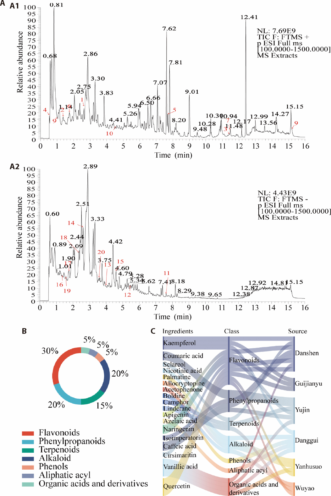
Figure 1 Identification of chemical constituents in XSD by UHPLC-MS/MS A: A1: XSD in ESI positive mode; A2: XSD in ESI negative mode. The red numbers in the mass spectrum represent 20 identified compounds. 1: kaempferol; 2: coumaric acid; 3: sclareol; 4: nicotinic acid; 5: palmatine; 6: allocryptopine; 7: acetophenone; 8: boldine; 9: camphor; 10: linderane; 11: apigenin; 12: kaempferol; 13: azelaic acid; 14: naringenin; 15: isoimperatorin; 16: coumaric acid; 17: caffeic acid; 18: cirsimaritin; 19: vanillic acid; 20: quercetin; B: metabolite class composition ring diagram; C: sankey diagram illustrating relationships between the ingredients and source herbs in XSD. XSD: Xuandan Sanjie decoction; UHPLC-MS/MS: ultra-high performance liquid chromatography coupled with tandem mass spectrometry; ESI: electrospray ionization.
Figure 1 Identification of chemical constituents in XSD by UHPLC-MS/MS A: A1: XSD in ESI positive mode; A2: XSD in ESI negative mode. The red numbers in the mass spectrum represent 20 identified compounds. 1: kaempferol; 2: coumaric acid; 3: sclareol; 4: nicotinic acid; 5: palmatine; 6: allocryptopine; 7: acetophenone; 8: boldine; 9: camphor; 10: linderane; 11: apigenin; 12: kaempferol; 13: azelaic acid; 14: naringenin; 15: isoimperatorin; 16: coumaric acid; 17: caffeic acid; 18: cirsimaritin; 19: vanillic acid; 20: quercetin; B: metabolite class composition ring diagram; C: sankey diagram illustrating relationships between the ingredients and source herbs in XSD. XSD: Xuandan Sanjie decoction; UHPLC-MS/MS: ultra-high performance liquid chromatography coupled with tandem mass spectrometry; ESI: electrospray ionization.
Table 1 Characterization of compounds of XSD by UPLC-MS/MS
| No. | Compound | Formula | mzmed | ms2Adduct | Relative content | Class |
|---|---|---|---|---|---|---|
| 1 | Kaempferol | C15H10O6 | 287.055 | [M+H]+ | 3860556.051 | Flavonoids |
| 2 | Coumaric acid | C9H8O3 | 147.043 | [M+H-H2O]+ | 42418783.33 | Phenylpropanoids |
| 3 | sclareol | C20H36O2 | 326.305 | [M+NH4]+ | 31763629.87 | Terpenoids |
| 4 | Nicotinic acid | C6H5NO2 | 124.039 | [M+H] | 87096072.94 | Alkaloid |
| 5 | Palmatine | C21H22NO4 | 352.153 | [M]+ | 1414855.134 | Alkaloid |
| 6 | Allocryptopine | C21H23NO5 | 370.165 | [M+H] | 20554792.16 | Alkaloid |
| 7 | Acetophenone | C8H8O | 121.064 | [M+H] | 4027023.846 | Phenols |
| 8 | Boldine | C19H21NO4 | 328.153 | [M+H]+ | 4970057868 | Alkaloid |
| 9 | Camphor | C10H16O | 153.127 | [M+H] | 1193613.001 | Terpenoids |
| 10 | Linderane | C15H16O4 | 261.111 | [M+H] | 103593915.9 | Terpenoids |
| 11 | Apigenin | C15H10O5 | 269.045 | [M-H]- | 2469278.777 | Flavonoids |
| 12 | Kaempferol | C15H10O6 | 285.042 | [M-H]- | 3370501.518 | Flavonoids |
| 13 | Azelaic acid | C9H16O4 | 187.097 | [M-H]- | 6112865.346 | Aliphatic acyl |
| 14 | Naringenin | C15H12O5 | 271.060 | [M-H] | 7626711.301 | Flavonoids |
| 15 | isoimperatorin | C16H14O4 | 269.082 | [M-H]- | 11272505.7 | Phenylpropanoids |
| 16 | Coumaric acid | C9H8O3 | 163.039 | [M-H]- | 12214940.45 | Phenylpropanoids |
| 17 | Caffeic acid | C9H8O4 | 179.034 | [M-H]- | 42289183.19 | Phenylpropanoids |
| 18 | cirsimaritin | C17H14O6 | 313.071 | [M-H]- | 55540661.17 | Flavonoids |
| 19 | Vanillic acid | C8H8O4 | 167.034 | [M-H]- | 67417507.56 | Cinnamic acid derivatives |
| 20 | Quercetin | C15H10O7 | 301.037 | [M-H]- | 3860556.051 | Flavonoids |
Table 1 Characterization of compounds of XSD by UPLC-MS/MS
| No. | Compound | Formula | mzmed | ms2Adduct | Relative content | Class |
|---|---|---|---|---|---|---|
| 1 | Kaempferol | C15H10O6 | 287.055 | [M+H]+ | 3860556.051 | Flavonoids |
| 2 | Coumaric acid | C9H8O3 | 147.043 | [M+H-H2O]+ | 42418783.33 | Phenylpropanoids |
| 3 | sclareol | C20H36O2 | 326.305 | [M+NH4]+ | 31763629.87 | Terpenoids |
| 4 | Nicotinic acid | C6H5NO2 | 124.039 | [M+H] | 87096072.94 | Alkaloid |
| 5 | Palmatine | C21H22NO4 | 352.153 | [M]+ | 1414855.134 | Alkaloid |
| 6 | Allocryptopine | C21H23NO5 | 370.165 | [M+H] | 20554792.16 | Alkaloid |
| 7 | Acetophenone | C8H8O | 121.064 | [M+H] | 4027023.846 | Phenols |
| 8 | Boldine | C19H21NO4 | 328.153 | [M+H]+ | 4970057868 | Alkaloid |
| 9 | Camphor | C10H16O | 153.127 | [M+H] | 1193613.001 | Terpenoids |
| 10 | Linderane | C15H16O4 | 261.111 | [M+H] | 103593915.9 | Terpenoids |
| 11 | Apigenin | C15H10O5 | 269.045 | [M-H]- | 2469278.777 | Flavonoids |
| 12 | Kaempferol | C15H10O6 | 285.042 | [M-H]- | 3370501.518 | Flavonoids |
| 13 | Azelaic acid | C9H16O4 | 187.097 | [M-H]- | 6112865.346 | Aliphatic acyl |
| 14 | Naringenin | C15H12O5 | 271.060 | [M-H] | 7626711.301 | Flavonoids |
| 15 | isoimperatorin | C16H14O4 | 269.082 | [M-H]- | 11272505.7 | Phenylpropanoids |
| 16 | Coumaric acid | C9H8O3 | 163.039 | [M-H]- | 12214940.45 | Phenylpropanoids |
| 17 | Caffeic acid | C9H8O4 | 179.034 | [M-H]- | 42289183.19 | Phenylpropanoids |
| 18 | cirsimaritin | C17H14O6 | 313.071 | [M-H]- | 55540661.17 | Flavonoids |
| 19 | Vanillic acid | C8H8O4 | 167.034 | [M-H]- | 67417507.56 | Cinnamic acid derivatives |
| 20 | Quercetin | C15H10O7 | 301.037 | [M-H]- | 3860556.051 | Flavonoids |

Figure 2 XSD inhibits angiogenesis by modulating VEGF and MMP9 secretion in vitro A: VEGF levels in ESC supernatants after 24 h of treatment; B: MMP9 levels in ESC supernatants after 24 h of treatment; C: C1-C7: tube formation assays of HUVECs treated with conditioned media from each group (× 40); C8-C14: representative images of endothelial tube formation analyzed by AngioTool; D: migration assays of HUVECs treated with conditioned media (× 100); E: invasion assays of HUVECs treated with conditioned media (× 100). C1, C8, D1, E1: Control group; C2, C9, D2, E2: E2 group; C3, C10, D3, E3: Vehicle group; C4, C11, D4, E4: Low dose XSD group; C5, C12, D5, E5: Middle dose XSD group; C6, C13, D6, E6: High dose XSD group; C7, C14, D7, E7: Mifepristone group. Control: ESCs treated with 200 μM CoCl2. E2: ESCs treated with 10 nM E2. Vehicle: ESCs treated with blank. XSD-L: ESCs treated with 2.5% (v/v) XSD-containing serum. XSD-M: ESCs treated with 5% (v/v) XSD-containing serum. XSD-H: ESCs treated with 10% (v/v) XSD-containing serum. Mifepristone: ESCs treated with 10 μM mifepristone. VEGF: vascular endothelial growth factor; MMP9: matrix metalloproteinase-9; ESCs: endometrial stromal cells; E2: Estrogen; XSD: Xuandan Sanjie decoction; ANOVA: analysis of variance. ANOVA tests were used among these groups. Data were presented as mean ± standard deviation (n = 3). Compared with the Control group, aP < 0.05; compared with the Vehicle group, bP > 0.05, cP < 0.05, dP < 0.01.
Figure 2 XSD inhibits angiogenesis by modulating VEGF and MMP9 secretion in vitro A: VEGF levels in ESC supernatants after 24 h of treatment; B: MMP9 levels in ESC supernatants after 24 h of treatment; C: C1-C7: tube formation assays of HUVECs treated with conditioned media from each group (× 40); C8-C14: representative images of endothelial tube formation analyzed by AngioTool; D: migration assays of HUVECs treated with conditioned media (× 100); E: invasion assays of HUVECs treated with conditioned media (× 100). C1, C8, D1, E1: Control group; C2, C9, D2, E2: E2 group; C3, C10, D3, E3: Vehicle group; C4, C11, D4, E4: Low dose XSD group; C5, C12, D5, E5: Middle dose XSD group; C6, C13, D6, E6: High dose XSD group; C7, C14, D7, E7: Mifepristone group. Control: ESCs treated with 200 μM CoCl2. E2: ESCs treated with 10 nM E2. Vehicle: ESCs treated with blank. XSD-L: ESCs treated with 2.5% (v/v) XSD-containing serum. XSD-M: ESCs treated with 5% (v/v) XSD-containing serum. XSD-H: ESCs treated with 10% (v/v) XSD-containing serum. Mifepristone: ESCs treated with 10 μM mifepristone. VEGF: vascular endothelial growth factor; MMP9: matrix metalloproteinase-9; ESCs: endometrial stromal cells; E2: Estrogen; XSD: Xuandan Sanjie decoction; ANOVA: analysis of variance. ANOVA tests were used among these groups. Data were presented as mean ± standard deviation (n = 3). Compared with the Control group, aP < 0.05; compared with the Vehicle group, bP > 0.05, cP < 0.05, dP < 0.01.
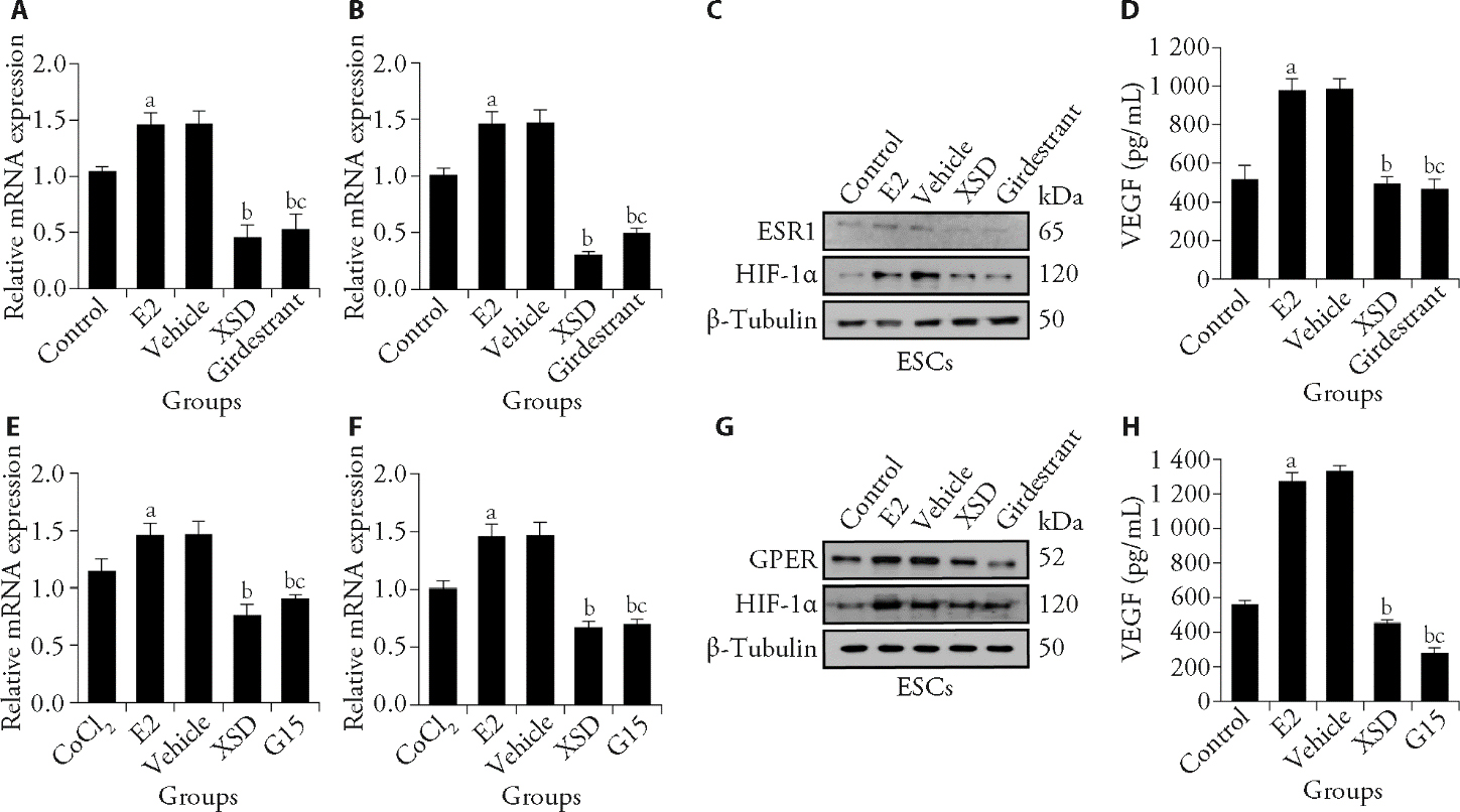
Figure 3 XSD regulates VEGF and MMP9 expression via ESR1/GPER-HIF-1α pathways A: qPCR analysis of HIF-1α mRNA in ESCs after 24 h; B: qPCR analysis of VEGF mRNA after 24 h; C: Western blot of ESR1 and HIF-1α at 24 h; D: VEGF levels in ESC supernatants (24 h); E: early (1 h) expression of HIF-1α mRNA; F: early (1 h) expression of VEGF mRNA; G: Western blot of GPER and HIF-1α at 1 h post-stimulation; H: VEGF secretion after 1 h assessed by ELISA. Group definitions: Control: 200 μM CoCl2; E2: 10 nM E2 under hypoxia; Vehicle: blank serum under hypoxia + E2; XSD: 10% (v/v) XSD serum under hypoxia + E2; Giredestrant: 10 nM Giredestrant under hypoxia + E2; G15: 100 nM G15 under hypoxia + E2. qPCR: quantitative reverse transcription polymerase chain reaction; ESCs: endometrial stromal cells; VEGF: vascular endothelial growth factor; MMP9: matrix metalloproteinase-9; CoCl2: cobalt (II) chloride; ESR1: estrogen receptor 1; HIF-1α: hypoxia inducible factor-1α; ELISA: enzyme-linked immunosorbent assay; GPER: G protein-coupled estrogen receptor; PI3K: phosphatidylinositol 3-kinase; p-PI3K: phospho-PI3K; AKT: protein kinase B; p-AKT: phospho-AKT; ERK: extracellular signal-regulated kinase; p-ERK: phospho-ERK; XSD: Xuandan Sanjie decoction; ANOVA: analysis of variance. ANOVA tests were used among these groups. Data were presented as mean ± standard deviation (n = 3). Compared with the Control group, aP < 0.05; compared with the Vehicle group, bP < 0.05; compared with the XSD group, cP > 0.05.
Figure 3 XSD regulates VEGF and MMP9 expression via ESR1/GPER-HIF-1α pathways A: qPCR analysis of HIF-1α mRNA in ESCs after 24 h; B: qPCR analysis of VEGF mRNA after 24 h; C: Western blot of ESR1 and HIF-1α at 24 h; D: VEGF levels in ESC supernatants (24 h); E: early (1 h) expression of HIF-1α mRNA; F: early (1 h) expression of VEGF mRNA; G: Western blot of GPER and HIF-1α at 1 h post-stimulation; H: VEGF secretion after 1 h assessed by ELISA. Group definitions: Control: 200 μM CoCl2; E2: 10 nM E2 under hypoxia; Vehicle: blank serum under hypoxia + E2; XSD: 10% (v/v) XSD serum under hypoxia + E2; Giredestrant: 10 nM Giredestrant under hypoxia + E2; G15: 100 nM G15 under hypoxia + E2. qPCR: quantitative reverse transcription polymerase chain reaction; ESCs: endometrial stromal cells; VEGF: vascular endothelial growth factor; MMP9: matrix metalloproteinase-9; CoCl2: cobalt (II) chloride; ESR1: estrogen receptor 1; HIF-1α: hypoxia inducible factor-1α; ELISA: enzyme-linked immunosorbent assay; GPER: G protein-coupled estrogen receptor; PI3K: phosphatidylinositol 3-kinase; p-PI3K: phospho-PI3K; AKT: protein kinase B; p-AKT: phospho-AKT; ERK: extracellular signal-regulated kinase; p-ERK: phospho-ERK; XSD: Xuandan Sanjie decoction; ANOVA: analysis of variance. ANOVA tests were used among these groups. Data were presented as mean ± standard deviation (n = 3). Compared with the Control group, aP < 0.05; compared with the Vehicle group, bP < 0.05; compared with the XSD group, cP > 0.05.
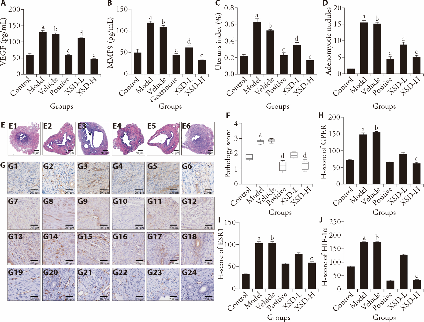
Figure 4 XSD inhibits angiogenesis in vivo in AM through ESR1/GPER-HIF-1α signaling A: VEGF levels in ectopic lesions from all groups (ELISA); B: MMP9 levels in ectopic lesions from all groups (ELISA); C: uterus index comparison; D: number of AM nodules; E: HE staining of uterine tissues (× 10); F: pathology scoring; G: immunohistochemistry staining of GPER, ESR1, HIF-1α, and CD31 (× 200); E1, G1, G7, G13, G19: Control group; E2, G2, G8, G14, G20: Model group; E3, G3, G9, G15, G21: Vehicle group; E4, G4, G10, G16, G22: Positive group; E5, G5, G11, G17, G23: XSD-L group; E6, G6, G12, G18, G24: XSD-H group. H: H-score quantification of GPER. I: H-score quantification of ESR1. J: H-score quantification of HIF-1α. Group definitions: Control: untreated mice; Model: saline (0.2 mL/d); Vehicle: distilled water (0.2 mL/d); Positive control (Mifepristone): 3.25 mg/kg bw; XSD-L: low dose (5.2 g/kg bw) XSD; XSD-H: high dose (10.4 g/kg bw) XSD. ESR1: estrogen receptor 1; GPER: G protein-coupled estrogen receptor; HIF-1α: hypoxia inducible factor-1α; VEGF: vascular endothelial growth factor; MMP9: matrix metalloproteinase-9; HE: hematoxylin and eosin; ELISA: enzyme-linked immunosorbent assay; AM: adenomyosis; XSD: Xuandan Sanjie decoction; ANOVA: analysis of variance. Statistical analysis was carried out using ANOVA tests among these groups. Data were presented as mean ± standard deviation (n = 8). Compared with the Control group, aP < 0.05; compared with the Model group, bP > 0.05, cP < 0.01, dP < 0.05.
Figure 4 XSD inhibits angiogenesis in vivo in AM through ESR1/GPER-HIF-1α signaling A: VEGF levels in ectopic lesions from all groups (ELISA); B: MMP9 levels in ectopic lesions from all groups (ELISA); C: uterus index comparison; D: number of AM nodules; E: HE staining of uterine tissues (× 10); F: pathology scoring; G: immunohistochemistry staining of GPER, ESR1, HIF-1α, and CD31 (× 200); E1, G1, G7, G13, G19: Control group; E2, G2, G8, G14, G20: Model group; E3, G3, G9, G15, G21: Vehicle group; E4, G4, G10, G16, G22: Positive group; E5, G5, G11, G17, G23: XSD-L group; E6, G6, G12, G18, G24: XSD-H group. H: H-score quantification of GPER. I: H-score quantification of ESR1. J: H-score quantification of HIF-1α. Group definitions: Control: untreated mice; Model: saline (0.2 mL/d); Vehicle: distilled water (0.2 mL/d); Positive control (Mifepristone): 3.25 mg/kg bw; XSD-L: low dose (5.2 g/kg bw) XSD; XSD-H: high dose (10.4 g/kg bw) XSD. ESR1: estrogen receptor 1; GPER: G protein-coupled estrogen receptor; HIF-1α: hypoxia inducible factor-1α; VEGF: vascular endothelial growth factor; MMP9: matrix metalloproteinase-9; HE: hematoxylin and eosin; ELISA: enzyme-linked immunosorbent assay; AM: adenomyosis; XSD: Xuandan Sanjie decoction; ANOVA: analysis of variance. Statistical analysis was carried out using ANOVA tests among these groups. Data were presented as mean ± standard deviation (n = 8). Compared with the Control group, aP < 0.05; compared with the Model group, bP > 0.05, cP < 0.01, dP < 0.05.
| [1] | Habiba M, Guo SW, Benagiano G. Adenomyosis and abnormal uterine bleeding: review of the evidence. Biomolecules 2024; 14: 616. |
| [2] | Li B, Shi K, Jing C, Xu L, Kong M, Ba M. Successful management of cerebral venous sinus thrombosis due to adenomyosis: case reports and literature review. Clin Neurol Neurosurg 2023; 229: 107726. |
| [3] | Moldassarina RS. Modern view on the diagnostics and treatment of adenomyosis. Arch Gynecol Obstet 2023; 308: 171-81. |
| [4] | Fan Y, Zhu S, Liang X. Conservative surgical and drug therapies for adenomyosis medicine. Reprod Biol 2022; 22: 100664. |
| [5] | Harmsen MJ, Wong CFC, Mijatovic V, et al. Role of angiogenesis in adenomyosis-associated abnormal uterine bleeding and subfertility: a systematic review. Hum Reprod Update 2019; 25: 646-70. |
| [6] | Harmsen MJ, Juffermans LJM, Kroon MO, Griffioen AW, Huirne JAF. Anti-angiogenic therapy as potential treatment for adenomyosis. Angiogenesis 2025; 28: 12. |
| [7] | Zhong S, Qian L, Tan Y. Xue Ping Tablets treat abnormal uterine bleeding via VEGF-ERK1/2 pathway. Heliyon 2024; 10: e30079. |
| [8] | Ru J, Li P, Wang J, et al. TCMSP: a database of systems pharmacology for drug discovery from herbal medicines. J Cheminform 2014; 6: 13. |
| [9] | Szklarczyk D, Gable AL, Nastou KC, et al. The STRING database in 2021:customizable protein-protein networks, and functional characterization of user-uploaded gene/measurement sets. Nucleic Acids Res 2020; 49: D605-12. |
| [10] | Zhang H, Zhou C, Zhang Z, et al. Integration of network pharmacology and experimental validation to explore the pharmacological mechanisms of Zhuanggu Busui formula against osteoporosis. Front Endocrinol (Lausanne) 2022; 12: 841668. |
| [11] | Gan L, Li Y, Chen Y, et al. Transcriptome analysis of eutopic endometrial stromal cells in women with adenomyosis by RNA-sequencing. Bioengineered 2022; 13: 12637-49. |
| [12] | Zhang YB, Wang X, Meister E, et al. The effects of CoCl2 on HIF-1α protein under experimental conditions of autoprogressive hypoxia using mouse models. Int J Mol Sci 2014; 15: 10999-1012. |
| [13] | Di Mattia M, Mauro A, Delle Monache S, et al. Hypoxia-mimetic CoCl2 agent enhances pro-angiogenic activities in ovine amniotic epithelial cells-derived conditioned medium. Cells 2022; 11: 461. |
| [14] | Muñoz-Sánchez J, Chánez-Cárdenas ME. The use of cobalt chloride as a chemical hypoxia model. J Appl Toxicol 2018; 39: 556-70. |
| [15] | Ye Q, Zhang J, Zhang C, et al. Endothelial PRMT5 plays a crucial role in angiogenesis after acute ischemic injury. JCI Insight 2022; 7: e152481. |
| [16] | Huang X, Pan Q, Mao Z, et al. Kaempferol inhibits interleukin-1β stimulated matrix metalloproteinases by suppressing the MAPK-associated ERK and P38 signaling pathways. Mol Med Rep 2018; 18: 2697-704. |
| [17] | Zhang J, Li H, Wang W, Li H. Assessing the anti-inflammatory effects of quercetin using network pharmacology and in vitro experiments. Exp Ther Med 2022; 23: 301. |
| [18] | Guo S, Zhang D, Lu X, et al. Hypoxia and its possible relationship with endometrial receptivity in adenomyosis: a preliminary study. Reprod Biol Endocrinol 2021; 19: 7. |
| [19] | Li B, Zhang Q, Sun J, Lai D. Human amniotic epithelial cells improve fertility in an intrauterine adhesion mouse model. Stem Cell Res Ther 2019; 10: 257. |
| [20] | Zhou S, Yi T, Liu R, et al. Proteomics identification of annexin A2 as a key mediator in the metastasis and proangiogenesis of endometrial cells in human adenomyosis. Mol Cell Proteomics 2012; 11: M112.017988. |
| [21] | Gui T, Liu M, Yao B, et al. TCF3 is epigenetically silenced by EZH2 and DNMT3B and functions as a tumor suppressor in endometrial cancer. Cell Death Differ 2021; 28: 3316-28. |
| [22] | Wang X, Xia X, Xu E, et al. Estrogen receptor beta prevents signet ring cell gastric carcinoma progression in young patients by inhibiting pseudopodia formation via the mTOR-Arpc1b/EVL signaling pathway. Front Cell Dev Biol 2021; 8: 592919. |
| [23] | Ahmed S, Grant KG, Edwards LE, et al. Data-driven modeling reconciles kinetics of ERK phosphorylation, localization, and activity states. Molecular Systems Biology 2014; 10: 718. |
| [24] | Filippi I, Carrarelli P, Luisi S, et al. Different expression of hypoxic and angiogenic factors in human endometriotic lesions. Reprod Sci 2016; 23: 492-7. |
| [25] | Hsiao KY, Lin SC, Wu MH, Tsai SJ. Pathological functions of hypoxia in endometriosis. Front Biosci (Elite Ed) 2015; 7: 309-21. |
| [26] | Tirpe AA, Gulei D, Ciortea SM, Crivii C, Berindan-Neagoe I. Hypoxia: overview on hypoxia-mediated mechanisms with a focus on the role of HIF genes. Int J Mol Sci 2019; 20: 6140. |
| [27] | Khater M, Greco F, Osborn HMI. Antiangiogenic activity of flavonoids: a systematic review and Meta-analysis. Molecules 2020; 25: 4712. |
| [28] | Ghosh R, Bhowmik A, Biswas S, et al. Natural flavonoid Orientin restricts 5-Fluorouracil induced cancer stem cells mediated angiogenesis by regulating HIF1α and VEGFA in colorectal cancer. Mol Med 2025; 31: 85. |
| [29] | Kong C, Jeong C, Choi J, Kim K, Jeong J. Antiangiogenic effects of p-coumaric acid in human endothelial cells. Phytother Res 2012; 27: 317-23. |
| [30] | Zhou J, Xie X, Tang H, Peng C, Peng F. The bioactivities of sclareol: a mini review. Front Pharmacol 2022; 13: 1014105. |
| [31] | Akhigbe RE, Afolabi OA, Adegbola CA, Akhigbe TM, Oyedokun PA, Afolabi OA. Comparison of the effectiveness of levonorgestrel intrauterine system and dienogest in the management of adenomyosis: a systematic review and Meta-analysis. Eur J Obstet Gynecol Reprod Biol 2024; 300: 230-9. |
| [32] | Kamba T, McDonald DM. Mechanisms of adverse effects of anti-VEGF therapy for cancer. Br J Cancer 2007; 96: 1788-95. |
| [33] | Wang L, Fei Y, Qu H, et al. Five years of safety profile of bevacizumab: an analysis of real-world pharmacovigilance and randomized clinical trials. J Pharm Health Care Sci 2024; 10: 1. |
| [34] | Moawad G, Youssef Y, Fruscalzo A, et al. The present and the future of medical therapies for adenomyosis: a narrative review. J Clin Med 2023; 12: 6130. |
| Viewed | ||||||
|
Full text |
|
|||||
|
Abstract |
|
|||||