
Journal of Traditional Chinese Medicine ›› 2026, Vol. 46 ›› Issue (5): 1139-1148.DOI: 10.19852/j.cnki.jtcm.2026.05.008
• Original Articles • Previous Articles Next Articles
Gandouling (肝豆灵) alleviates Wilson disease-associated hepatic fibrosis through inhibiting DNA methyltransferase 3 alpha/dynamin-related protein 1 mediated mitochondrial fission
XUAN Qiaoyu, HUA Daiping, SUN Lanting, GE Shufan, YANG Wenming, WANG Han( )
Department of Neurology ,the First Affiliated Hospital of Anhui University of Chinese Medicine Hefei 230031, China
-
Received:2025-06-11Accepted:2026-02-07Online:2026-10-15Published:2026-10-09 -
Contact:Prof. WANG Han, Department of Neurology, the First Affiliated Hospital of Anhui University of Chinese Medicine, Hefei 230031, China. neuwhah@126.com,Telephone: +86-18356136585 -
About author:XUAN Qiaoyua and HUA Daipinga are co-first authors and contributed equally to this work
-
Supported by:Regional Innovation and Development Joint Fund of the National Natural Science Foundation of China: Study on the Mechanism of Xin'an Medical Guben Peiyuan Formula in Treating Wilson Disease Based on the Gut-liver Axis and Gut-brain Axis Pathways(U22A20366);Anhui Provincial Natural Science Foundation: Study on the Mechanism of Gandouling Against Hepatic Fibrosis in Wilson Disease via Regulating Hepatic Stellate Cell Autophagy by lncRNA Meg3(2208085MH266);Anhui Provincial Special Project for the Development of Traditional Chinese Medicine: High-level Talent Inheritance Program(Anhui TCM Development Document No. 1 [2024])

Cite this article
XUAN Qiaoyu, HUA Daiping, SUN Lanting, GE Shufan, YANG Wenming, WANG Han. Gandouling (肝豆灵) alleviates Wilson disease-associated hepatic fibrosis through inhibiting DNA methyltransferase 3 alpha/dynamin-related protein 1 mediated mitochondrial fission[J]. Journal of Traditional Chinese Medicine, 2026, 46(5): 1139-1148.
share this article
Table 1 Top 5 active component chemical compositions of GDL
| MOL ID | Degree | Molecule name | Molecular formula | CAS | Source |
|---|---|---|---|---|---|
| MOL002293 | 71 | Sennoside D | C42H40O19 | 37271-17-3 | DH |
| MOL000006 | 66 | Luteolin | C15H10O6 | 491-70-3 | DS, JXT |
| MOL000296 | 62 | Hederagenin | C30H48O4 | 465-99-6 | EZ, JXT |
| MOL013352 | 56 | Obacunone | C26H30O7 | 751-03-1 | HL |
| MOL000493 | 54 | campesterol | C28H48O | 474-62-4 | JH, JXT |
Table 1 Top 5 active component chemical compositions of GDL
| MOL ID | Degree | Molecule name | Molecular formula | CAS | Source |
|---|---|---|---|---|---|
| MOL002293 | 71 | Sennoside D | C42H40O19 | 37271-17-3 | DH |
| MOL000006 | 66 | Luteolin | C15H10O6 | 491-70-3 | DS, JXT |
| MOL000296 | 62 | Hederagenin | C30H48O4 | 465-99-6 | EZ, JXT |
| MOL013352 | 56 | Obacunone | C26H30O7 | 751-03-1 | HL |
| MOL000493 | 54 | campesterol | C28H48O | 474-62-4 | JH, JXT |
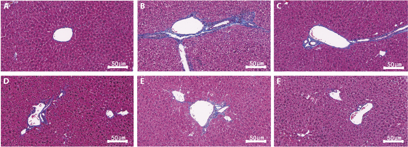
Figure 1 Masson staining for detection of fibrosis in liver tissue A: Control; B: Model; C: GDL-L; D: GDL-M; E: GDL-H; F: PCA (magnification × 200, scale bar = 50 μm). Control: equal-volume distilled water. Model: modeling plus intragastric administration of distilled water. GDL-L/M/H: 0.24, 0.48, 0.96 g/kg GDL, respectively. PCA: 90 mg/kg PCA. GDL: Gandouling; GDL-L: low-dose GDL group; GDL-M: medium-dose GDL group; GDL-H: high-dose GDL group; PCA: penicillamine.
Figure 1 Masson staining for detection of fibrosis in liver tissue A: Control; B: Model; C: GDL-L; D: GDL-M; E: GDL-H; F: PCA (magnification × 200, scale bar = 50 μm). Control: equal-volume distilled water. Model: modeling plus intragastric administration of distilled water. GDL-L/M/H: 0.24, 0.48, 0.96 g/kg GDL, respectively. PCA: 90 mg/kg PCA. GDL: Gandouling; GDL-L: low-dose GDL group; GDL-M: medium-dose GDL group; GDL-H: high-dose GDL group; PCA: penicillamine.
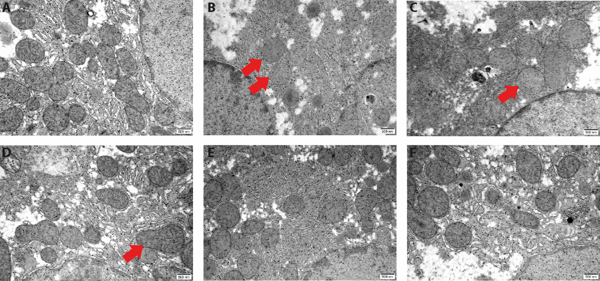
Figure 2 GDL-associated effects on mitochondrial changes in WD model rats A: Control; B: Model; C: GDL-L; D: GDL-M; E: GDL-H; F: PCA (mitochondria are indicated by red arrows. × 25 000 magnification, scale bar: 500 nm). Control: equal-volume distilled water; Model: modeling plus intragastric administration of distilled water; GDL-L/M/H: 0.24, 0.48, 0.96 g/kg GDL, respectively; PCA: 90 mg/kg PCA. GDL: Gandouling; GDL-L: low-dose GDL group; GDL-M: medium-dose GDL group; GDL-H: high-dose GDL group; PCA: penicillamine.
Figure 2 GDL-associated effects on mitochondrial changes in WD model rats A: Control; B: Model; C: GDL-L; D: GDL-M; E: GDL-H; F: PCA (mitochondria are indicated by red arrows. × 25 000 magnification, scale bar: 500 nm). Control: equal-volume distilled water; Model: modeling plus intragastric administration of distilled water; GDL-L/M/H: 0.24, 0.48, 0.96 g/kg GDL, respectively; PCA: 90 mg/kg PCA. GDL: Gandouling; GDL-L: low-dose GDL group; GDL-M: medium-dose GDL group; GDL-H: high-dose GDL group; PCA: penicillamine.
Table 2 Comparisons of serum AST, ALT, HA, LN, PC-III, and C-IV in rats among different groups ($\stackrel{-}{\mathit{x}}\mathrm{ }$± s)
| Groups | n | AST | ALT | HA | LN | PC-III | C-IV |
|---|---|---|---|---|---|---|---|
| Control | 6 | 31.74±9.20 | 29.44±3.66 | 15.78±2.60 | 201.53±14.97 | 3129.93±155.76 | 5.57±0.33 |
| Model | 6 | 148.04±16.23a | 101.80±10.29a | 67.71±3.27a | 637.18±11.08a | 7336.30±193.59a | 19.38±0.51a |
| GDL-L | 6 | 115.36±10.07b | 95.07±3.34 | 60.47±0.58b | 559.79±2.96b | 6672.49±93.04b | 17.62±0.55b |
| GDL-M | 6 | 100.04±8.99b | 89.00±7.74b | 53.50±2.11b | 480.62±15.72b | 5297.46±179.90b | 14.02±0.63b |
| GDL-H | 6 | 59.20±4.02b | 59.35±3.21b | 31.88±2.00b | 413.65±24.58b | 4219.81±164.06b | 10.45±0.25b |
| PCA | 6 | 62.46±6.82b | 62.12±4.82b | 29.94±1.71b | 424.93±5.13b | 4352.91±132.90b | 11.28±0.76b |
| F value | 110.88 | 121.37 | 508.84 | 652.01 | 620.59 | 536.49 | |
| P value | < 0.001 | < 0.001 | < 0.001 | < 0.001 | < 0.001 | < 0.001 |
Table 2 Comparisons of serum AST, ALT, HA, LN, PC-III, and C-IV in rats among different groups ($\stackrel{-}{\mathit{x}}\mathrm{ }$± s)
| Groups | n | AST | ALT | HA | LN | PC-III | C-IV |
|---|---|---|---|---|---|---|---|
| Control | 6 | 31.74±9.20 | 29.44±3.66 | 15.78±2.60 | 201.53±14.97 | 3129.93±155.76 | 5.57±0.33 |
| Model | 6 | 148.04±16.23a | 101.80±10.29a | 67.71±3.27a | 637.18±11.08a | 7336.30±193.59a | 19.38±0.51a |
| GDL-L | 6 | 115.36±10.07b | 95.07±3.34 | 60.47±0.58b | 559.79±2.96b | 6672.49±93.04b | 17.62±0.55b |
| GDL-M | 6 | 100.04±8.99b | 89.00±7.74b | 53.50±2.11b | 480.62±15.72b | 5297.46±179.90b | 14.02±0.63b |
| GDL-H | 6 | 59.20±4.02b | 59.35±3.21b | 31.88±2.00b | 413.65±24.58b | 4219.81±164.06b | 10.45±0.25b |
| PCA | 6 | 62.46±6.82b | 62.12±4.82b | 29.94±1.71b | 424.93±5.13b | 4352.91±132.90b | 11.28±0.76b |
| F value | 110.88 | 121.37 | 508.84 | 652.01 | 620.59 | 536.49 | |
| P value | < 0.001 | < 0.001 | < 0.001 | < 0.001 | < 0.001 | < 0.001 |
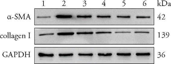
Figure 3 Western blotting analysis of α-SMA and collagen I protein expression 1: Control; 2: Model; 3: GDL-L; 4: GDL-M; 5: GDL-H; 6: PCA. Control: equal-volume distilled water; Model: modeling plus intragastric administration of distilled water; GDL-L/M/H: 0.24, 0.48, 0.96 g/kg GDL, respectively; PCA: 90 mg/kg PCA. GDL: Gandouling; GDL-L: low-dose GDL group; GDL-M: medium-dose GDL group; GDL-H: high-dose GDL group; PCA: penicillamine; α-SMA: α-smooth muscle actin; collagen Ⅰ: type Ⅰ collagen; GAPDH: glyceraldehyde-3-phosphate dehydrogenase.
Figure 3 Western blotting analysis of α-SMA and collagen I protein expression 1: Control; 2: Model; 3: GDL-L; 4: GDL-M; 5: GDL-H; 6: PCA. Control: equal-volume distilled water; Model: modeling plus intragastric administration of distilled water; GDL-L/M/H: 0.24, 0.48, 0.96 g/kg GDL, respectively; PCA: 90 mg/kg PCA. GDL: Gandouling; GDL-L: low-dose GDL group; GDL-M: medium-dose GDL group; GDL-H: high-dose GDL group; PCA: penicillamine; α-SMA: α-smooth muscle actin; collagen Ⅰ: type Ⅰ collagen; GAPDH: glyceraldehyde-3-phosphate dehydrogenase.
Table 3 Relative protein expression levels of α-SMA and collagen I in different groups ($\stackrel{-}{\mathit{x}}\mathrm{ }$± s)
| Groups | n | α-SMA | collagen I |
|---|---|---|---|
| Control | 3 | 0.140±0.022 | 0.212±0.049 |
| Model | 3 | 1.102±0.028a | 0.763±0.042a |
| GDL-L | 3 | 0.990±0.049 | 0.654±0.054 |
| GDL-M | 3 | 0.736±0.075b | 0.549±0.045b |
| GDL-H | 3 | 0.541±0.050b | 0.356±0.072b |
| PCA | 3 | 0.503±0.043b | 0.322±0.088b |
| F value | 162.401 | 36.928 | |
| P value | < 0.001 | < 0.001 |
Table 3 Relative protein expression levels of α-SMA and collagen I in different groups ($\stackrel{-}{\mathit{x}}\mathrm{ }$± s)
| Groups | n | α-SMA | collagen I |
|---|---|---|---|
| Control | 3 | 0.140±0.022 | 0.212±0.049 |
| Model | 3 | 1.102±0.028a | 0.763±0.042a |
| GDL-L | 3 | 0.990±0.049 | 0.654±0.054 |
| GDL-M | 3 | 0.736±0.075b | 0.549±0.045b |
| GDL-H | 3 | 0.541±0.050b | 0.356±0.072b |
| PCA | 3 | 0.503±0.043b | 0.322±0.088b |
| F value | 162.401 | 36.928 | |
| P value | < 0.001 | < 0.001 |
Table 4 Relative mRNA expression levels of Drp1 and DNMT3A in different groups ($\stackrel{-}{\mathit{x}}\mathrm{ }$± s)
| Groups | n | Drp1 | DNMT3A |
|---|---|---|---|
| Control | 6 | 1.00±0.07 | 1.00±0.04 |
| Model | 6 | 2.88±0.14a | 1.62±0.04a |
| GDL-L | 6 | 1.87±0.04b | 1.43±0.03b |
| GDL-M | 6 | 1.50±0.03b | 1.34±0.04b |
| GDL-H | 6 | 1.33±0.08b | 1.16±0.05b |
| PCA | 6 | 1.30±0.07b | 1.16±0.05b |
| F value | 405.93 | 175.26 | |
| P value | < 0.001 | < 0.001 |
Table 4 Relative mRNA expression levels of Drp1 and DNMT3A in different groups ($\stackrel{-}{\mathit{x}}\mathrm{ }$± s)
| Groups | n | Drp1 | DNMT3A |
|---|---|---|---|
| Control | 6 | 1.00±0.07 | 1.00±0.04 |
| Model | 6 | 2.88±0.14a | 1.62±0.04a |
| GDL-L | 6 | 1.87±0.04b | 1.43±0.03b |
| GDL-M | 6 | 1.50±0.03b | 1.34±0.04b |
| GDL-H | 6 | 1.33±0.08b | 1.16±0.05b |
| PCA | 6 | 1.30±0.07b | 1.16±0.05b |
| F value | 405.93 | 175.26 | |
| P value | < 0.001 | < 0.001 |
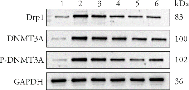
Figure 4 Western blotting analysis of Drp1, DNMT3A and p-DNMT3A protein expression 1: Control; 2: Model; 3: GDL-L; 4: GDL-M; 5: GDL-H; 6: PCA. Control: equal-volume distilled water; Model: modeling plus intragastric administration of distilled water; GDL-L/M/H: 0.24, 0.48, 0.96 g/kg GDL, respectively; PCA: 90 mg/kg PCA. GDL: Gandouling; GDL-L: low-dose GDL group; GDL-M: medium-dose GDL group; GDL-H: high-dose GDL group; PCA: penicillamine; Drp1: dynamin-related protein 1; DNMT3A: DNA methyltransferase 3 alpha; p-DNMT3A: phosphorylated DNMT3A; GAPDH: glyceraldehyde-3-phosphate dehydrogenase.
Figure 4 Western blotting analysis of Drp1, DNMT3A and p-DNMT3A protein expression 1: Control; 2: Model; 3: GDL-L; 4: GDL-M; 5: GDL-H; 6: PCA. Control: equal-volume distilled water; Model: modeling plus intragastric administration of distilled water; GDL-L/M/H: 0.24, 0.48, 0.96 g/kg GDL, respectively; PCA: 90 mg/kg PCA. GDL: Gandouling; GDL-L: low-dose GDL group; GDL-M: medium-dose GDL group; GDL-H: high-dose GDL group; PCA: penicillamine; Drp1: dynamin-related protein 1; DNMT3A: DNA methyltransferase 3 alpha; p-DNMT3A: phosphorylated DNMT3A; GAPDH: glyceraldehyde-3-phosphate dehydrogenase.
Table 5 Relative protein expression levels of Drp1, DNMT3A and p-DNMT3A in different groups ($\stackrel{-}{\mathit{x}}\mathrm{ }$± s)
| Group | n | Drp1 | DNMT3A | p-DNMT3A |
|---|---|---|---|---|
| Control | 3 | 0.214±0.014 | 0.205±0.073 | 0.078±0.024 |
| Model | 3 | 0.810±0.012a | 0.850±0.064a | 0.823±0.025a |
| GDL-L | 3 | 0.699±0.042 | 0.745±0.051 | 0.723±0.050 |
| GDL-M | 3 | 0.581±0.059b | 0.613±0.081b | 0.631±0.037b |
| GDL-H | 3 | 0.476±0.070b | 0.470±0.092b | 0.416±0.107b |
| PCA | 3 | 0.411±0.027b | 0.463±0.079b | 0.459±0.058b |
| F value | 72.501 | 28.686 | 64.508 | |
| P value | < 0.001 | < 0.001 | < 0.001 |
Table 5 Relative protein expression levels of Drp1, DNMT3A and p-DNMT3A in different groups ($\stackrel{-}{\mathit{x}}\mathrm{ }$± s)
| Group | n | Drp1 | DNMT3A | p-DNMT3A |
|---|---|---|---|---|
| Control | 3 | 0.214±0.014 | 0.205±0.073 | 0.078±0.024 |
| Model | 3 | 0.810±0.012a | 0.850±0.064a | 0.823±0.025a |
| GDL-L | 3 | 0.699±0.042 | 0.745±0.051 | 0.723±0.050 |
| GDL-M | 3 | 0.581±0.059b | 0.613±0.081b | 0.631±0.037b |
| GDL-H | 3 | 0.476±0.070b | 0.470±0.092b | 0.416±0.107b |
| PCA | 3 | 0.411±0.027b | 0.463±0.079b | 0.459±0.058b |
| F value | 72.501 | 28.686 | 64.508 | |
| P value | < 0.001 | < 0.001 | < 0.001 |
| [1] | Czlonkowska A, Litwin T, Dusek P, et al. Wilson disease. Nat Rev Dis Primers 2018; 4: 21-65. |
| [2] | Wan LY, Peng H, Ni YR, et al. The miR-23b/27b/24-1 cluster inhibits hepatic fibrosis by inactivating hepatic stellate cells. Cell Mol Gastroenterol Hepatol 2022; 13: 1393-412. |
| [3] | Zong Y, Li H, Liao P, et al. Mitochondrial dysfunction: mechanisms and advances in therapy. Signal Transduct Target Ther 2024; 9: 124-53. |
| [4] | Hua FZ, Zhu H, Yu W, et al. β-arrestin 1 regulates astrocytic reactivity via Drp1-dependent mitochondrial fission: implications in postoperative delirium. J Neuroinflammation 2023; 20: 113-38. |
| [5] | Zhou YN, Long D, Zhao Y, et al. Oxidative stress-mediated mitochondrial fission promotes hepatic stellate cell activation via stimulating oxidative phosphorylation. Cell Death Dis 2022; 13: 689-704. |
| [6] | Hasan P, Saotome M, Ikoma T, et al. Mitochondrial fission protein, dynamin-related protein 1, contributes to the promotion of hypertensive cardiac hypertrophy and fibrosis in Dahl-salt sensitive rats. J Mol Cell Cardiol 2018; 121: 103-6. |
| [7] | Wang YT, Lu MQ, Xiong LP, et al. Drp1-mediated mitochondrial fission promotes renal fibroblast activation and fibrogenesis. Cell Death Dis 2020; 11: 29-43. |
| [8] | Li DM, Guo XS, Zhao WY, Jingyu JY, Xia C, Yu GY. Genome-wide DNA methylation dynamics in carbon tetrachloride-induced mice liver fibrosis. Iran J Basic Med Sci 2023; 26: 85-92. |
| [9] | Yang JJ, Wang J, Yang Y, et al. Epigenetic control of mitochondrial fission enables hepatic stellate cells activation in liver fibrosis via PGC-1α-Drp1 pathway. Mitochondrion 2022; 66: 38-50. |
| [10] | Li XY, Wang H, Sun LT, et al. Mechanism of GanDouLing in improving liver fibrosis in Wilson disease based on network pharmacology and experimental verification. Hainan Yi Xue Yuan Xue Bao 2022; 28: 1644-50. |
| [11] | Cheng CL, Wang Q, Huang YR, et al. Gandouling inhibits hepatic fibrosis in Wilson's disease through Wnt-1/β-catenin signaling pathway. J Ethnopharmacol 2023; 311: 116445-55. |
| [12] | Zhao CL, Chen J, Tian LW, et al. Gandouling ameliorates liver injury in Wilson's disease through the inhibition of ferroptosis by regulating the HSF1/HSPB1 pathway. J Cell Mol Med 2024; 28: e70018-31. |
| [13] | Li S, Zhang B. Traditional Chinese Medicine network pharmacology: theory, methodology and application. Chin J Nat Med 2013; 11: 110-20. |
| [14] | Zhang MH, Zhang JL, Wu XF, et al. Study on the mechanism of baicalin on copper-loaded hepatolenticular degeneration in rats through copper metabolism. Zhong Yao Xin Yao Yu Lin Chuang Yao Li 2023; 34: 1211-8. |
| [15] | Xu SY, Bian RL, Chen X. Pharmacological experimental methodology. 3rd ed. Beijing: People's Medical Publishing House, 2002: 179-89. |
| [16] | Rao ZH, Yang WM, Yang YL, et al. Pathogenesis explanation of hepatolenticular degeneration along the "liver-kidney-brain" axis and differentiation and treatment strategies of Traditional Chinese Medicine. Beijing Zhong Yi Yao Da Xue Xue Bao 2025; 48: 1270-7. |
| [17] | Ding YF, Yang WM, Cheng Y, et al. Relative exchangeable copper, a high-quality biomarker for differentiation of Traditional Chinese Medicine syndrome in Wilson's disease. J Tradit Chin Med 2024; 44: 1017-23. |
| [18] | Zhang Y, Wang MX, Zhang J, et al. Clinical efficacy of Gandouling combined with short-term decoppering on hepatic fibrosis of Wilson's disease based on transient elastography. Zhong Guo Shi Yan Fang Ji Xue Za Zhi 2022; 28: 123-9. |
| [19] | Wang H, Hua DP, Yin X, Xuan QY, Sun LT, Yang WM. Exploration on the action mechanism of Gandouling in the treatment of Wilson disease liver fibrosis from the regulation of cell autophagy pathway by LncRNA Meg3. Zhong Hua Zhong Yi Yao Za Zhi 2025; 40: 2574-9. |
| [20] | Zhang J, Bao YC, Xie DJ, et al. Effect of Gandouling on serum metabolomics of liver fibrosis in copper-loaded rats. Zhong Yi Za Zhi 2014; 55: 232-7. |
| [21] | Liao XM, Ruan XX, Wu XB, Deng ZJ, Qin SY, Jiang HX. Identification of Timm13 protein translocase of the mitochondrial inner membrane as a potential mediator of liver fibrosis based on bioinformatics and experimental verification. J Transl Med 2023; 21: 188-99. |
| [22] | Yang Y, Hao WJ, Yang YL, et al. Chemical profiling and quantification of potential bioactive components in Gandouling pill by ultra-high performance liquid chromatography coupled with diode array detector/quadruple-qrbitrap mass spectrometry. Molecules 2022; 27: 8247-64. |
| [23] | Liu CC, Liu Q, Nian MN, et al. Identification and quantitative analysis of the chemical constituents of Gandouling tablets using ultra-high-performance liquid chromatography with quadrupole time-of-flight mass spectrometry. J Sep Sci 2023; 46: e2300060-85. |
| [24] | Zhu H, Zhao HZ, Xu SB, et al. Sennoside A alleviates inflammatory responses by inhibiting the hypermethylation of SOCS1 in CCl(4)-induced liver fibrosis. Pharmacol Res 2021; 174: 105926-38. |
| [25] | Batudeligen, Han ZQ, Chen HM, et al. Luteolin alleviates liver fibrosis in rat hepatic stellate cell HSC-T6: a proteomic analysis. Drug Des Devel Ther 2023; 17: 1819-29. |
| [26] | Xu J, Wang XD, Zhang HY, et al. Synthesis of triterpenoid derivatives and their anti-tumor and anti-hepatic fibrosis activities. Nat Prod Res 2020; 34: 766-72. |
| [27] | Yang GH, Yang W, Jiang HR, Yi Q, Ma W. Hederagenin inhibits high glucose-induced fibrosis in human renal cells by suppression of NLRP3 inflammasome activation through reducing cathepsin B expression. Chem Biol Drug Des 2023; 102: 1409-20. |
| [28] | Yang CY, Geng XY, Wan GG, et al. Transcriptomic and proteomic investigation of the ameliorative effect of total polyphenolic glycoside extract on hepatic fibrosis in Lamiophlomis rotata Kudo via the AGE/RAGE pathway. J Ethnopharmacol 2024; 324: 117720-35. |
| [29] | Zhang TT, Wang Y, Zhang XW, Yang KY, Miao XQ, Zhao GH. MiR-200c-3p regulates DUSP1/MAPK pathway in the nonalcoholic fatty liver after laparoscopic sleeve gastrectomy. Front Endocrinol 2022; 13: 792439-50. |
| [30] | Cingolani F, Czaja MJ. Oxidized albumin-A trojan horse for p38 MAPK-mediated inflammation in decompensated cirrhosis. Hepatology 2018; 68: 1678-80. |
| [31] | Xiu AY, Ding Q, Li Z, Zhang CQ. Doxazosin attenuates liver fibrosis by inhibiting autophagy in hepatic stellate cells via activation of the PI3K/Akt/mTOR signaling pathway. Drug Des Devel Ther 2021; 15: 3643-59. |
| [32] | Ye Q, Liu Y, Zhang GJ, et al. Deficiency of gluconeogenic enzyme PCK1 promotes metabolic-associated fatty liver disease through PI3K/AKT/PDGF axis activation in male mice. Nat Commun 2023; 14: 1402-21. |
| [33] | Zhang LZ, Zhang YH, Chang XX, Zhang XY. Imbalance in mitochondrial dynamics induced by low PGC-1α expression contributes to hepatocyte EMT and liver fibrosis. Cell Death Dis 2020; 11: 226-38. |
| [34] | Wang J, Yang Y, Sun F, et al. ALKBH 5 attenuates mitochondrial fission and ameliorates liver fibrosis by reducing Drp1 methylation. Pharmacol Res 2023; 187: 106608-25. |
| [35] | Shan SL, Liu ZD, Wang SA, et al. Drp1-mediated mitochondrial fission promotes carbon tetrachloride-induced hepatic fibrogenesis in mice. Toxicol Res 2022; 11: 486-97. |
| [36] | Zhao D, Gao Y, Su Y, et al. Oroxylin A regulates cGAS DNA hypermethylation induced by methionine metabolism to promote HSC senescence. Pharmacol Res 2023; 187: 106590-606. |
| [37] | Page A, Paoli P, Moran Salvador E, White S, French J, Mann J. Hepatic stellate cell transdifferentiation involves genome-wide remodeling of the DNA methylation landscape. J Hepatol 2016; 64: 661-73. |
| [38] | Challen GA, Sun DQ, Mayle A, et al. Dnmt3a and Dnmt3b have overlapping and distinct functions in hematopoietic stem cells. Cell Stem Cell 2014; 15: 350-64. |
| [39] | Zha YF, Li YY, Ge ZW, et al. ADAMTS8 promotes cardiac fibrosis partly through activating EGFR dependent pathway. Front Cardiovasc Med 2022; 9: 797137-55. |
| [40] | Yao XD, Jing XZ, Guo JC, et al. Icariin protects bone marrow mesenchymal stem cells against iron overload induced dysfunction through mitochondrial fusion and fission, PI3K/AKT/mTOR and MAPK pathways. Front Pharmacol 2019; 10: 163-77. |
| [41] | Rinaldi L, Avgustinova A, Martín M, et al. Loss of Dnmt3a and Dnmt3b does not affect epidermal homeostasis but promotes squamous transformation through PPAR-γ. eLife 2017; 6: e21697-722. |
| [42] | Yang S, Cheng M, Zhang SN, et al. DNMT3A facilitates breast cancer progression via regulating ADAMTS8 mediated EGFR-MEK-ERK activation. PLoS One 2025; 20: e0321889-906. |
| [43] | He D, Wang X, Zhang Y, Zhao J, Han R, Dong Y. DNMT3A/3B overexpression might be correlated with poor patient survival, hypermethylation and low expression of ESR1/PGR in endometrioid carcinoma: an analysis of The Cancer Genome Atlas. Chin Med J 2019; 132: 161-70. |
| Viewed | ||||||
|
Full text |
|
|||||
|
Abstract |
|
|||||