
Journal of Traditional Chinese Medicine ›› 2026, Vol. 46 ›› Issue (5): 1117-1126.DOI: 10.19852/j.cnki.jtcm.2026.05.006
• Original Articles • Previous Articles Next Articles
Grain-sized moxibustion attenuates renal dysfunction and inflammation in naturally aging mice via the phosphatidylinositol 3-kinase/protein kinase B/nuclear factor-κB pathway
XU Hui1,2, QI Li3, QIAN Yutong1, LIU Chang1, LI Zhenchen3, MENG Qi3, LIU Shimin1, JIAO Danli1( ), ZHAO Chen1()
- 1
School of Acupuncture-moxibustion and Tuina ,Shanghai University of Traditional Chinese Medicine Shanghai 201203, China
2Department of TCM ,Taizhou Second People's Hospital Taizhou 225500, China
3Institute of Interdisciplinary Integrative Medicine Research ,Shanghai University of Traditional Chinese Medicine Shanghai 201203, China
-
Received:2025-05-29Accepted:2025-11-05Online:2026-10-15Published:2026-10-09 -
Contact:Prof. ZHAO Chen, School of Acupuncture-moxibustion and Tuina, Shanghai University of Traditional Chinese Medicine, Shanghai 201203, China. ztzhaochen@126.com,Telephone: +86-13764276086; Prof. JIAO Danli, School of Acupuncture-moxibustion and Tuina, Shanghai University of Traditional Chinese Medicine, Shanghai 201203, China. 937095417@qq.com,Telephone: +86-18616124659 -
Supported by:Three-year Action Plan of the National Health and Family Planning Commission: Study on the Effect and Mechanism of Acupuncture and Moxibustion in Regulating Senescence-associated Secretory Phenotype to Alleviate Renal Fibrosis via Wnt family Member 9a /β-catenin Signaling Pathway(E2-ED19005);the Budget for the Project of Shanghai University of Traditional Chinese Medicine: Clinical Study of Eight-Shu Moxibustion in the Treatment of Early Diabetic Nephropathy(2021LK027);the Project of Clinical Research of the Health Industry of the Shanghai Municipal Health Commission(202340167)

Cite this article
XU Hui, QI Li, QIAN Yutong, LIU Chang, LI Zhenchen, MENG Qi, LIU Shimin, JIAO Danli, ZHAO Chen. Grain-sized moxibustion attenuates renal dysfunction and inflammation in naturally aging mice via the phosphatidylinositol 3-kinase/protein kinase B/nuclear factor-κB pathway[J]. Journal of Traditional Chinese Medicine, 2026, 46(5): 1117-1126.
share this article

Figure 1 GS-Moxi decreases age-associated renal dysfunction and histopathological morphology A: schematic diagram of the overall animal experimental design; B: representative PAS staining micrographs of mouse renal tissues (scale bar = 50 μm); B1: Young group; B2: Old group; B3: GS-Moxi group; B4: semiquantitative statistical scoring of renal tubular injury; C: representative Masson’s trichrome staining micrographs of mouse renal tissues (scale bar = 50 μm); C1: Young group; C2: Old group; C3: GS-Moxi group; C4: semiquantitative statistical scoring of renal interstitial fibrosis; D: relative mRNA expression of TGF-β1 detected by qPCR; E1: SCr concentration; E2: BUN concentration. Young group: untreated young mice without acupoint stimulation; Old group: naturally aged mice with spontaneous senescent renal injury; GS-Moxi group: aged mice treated with grain-sized moxibustion at Zusanli (ST36) acupoint for 12 consecutive weeks. GS-Moxi: grain-sized moxibustion; PAS: periodic acid-Schiff; qPCR: quantitative real-time polymerase chain reaction; TGF-β1: transforming growth factor beta 1; SCr: serum creatinine; BUN: blood urea nitrogen. Statistical analyses were measured by one-way analysis, and least significance difference test was performed for inter-group comparisons. All data were presented as mean ± standard deviation (n = 6). Compared with the young group, aP < 0.01, cP < 0.05; compared with the old group, bP < 0.01, dP < 0.05.
Figure 1 GS-Moxi decreases age-associated renal dysfunction and histopathological morphology A: schematic diagram of the overall animal experimental design; B: representative PAS staining micrographs of mouse renal tissues (scale bar = 50 μm); B1: Young group; B2: Old group; B3: GS-Moxi group; B4: semiquantitative statistical scoring of renal tubular injury; C: representative Masson’s trichrome staining micrographs of mouse renal tissues (scale bar = 50 μm); C1: Young group; C2: Old group; C3: GS-Moxi group; C4: semiquantitative statistical scoring of renal interstitial fibrosis; D: relative mRNA expression of TGF-β1 detected by qPCR; E1: SCr concentration; E2: BUN concentration. Young group: untreated young mice without acupoint stimulation; Old group: naturally aged mice with spontaneous senescent renal injury; GS-Moxi group: aged mice treated with grain-sized moxibustion at Zusanli (ST36) acupoint for 12 consecutive weeks. GS-Moxi: grain-sized moxibustion; PAS: periodic acid-Schiff; qPCR: quantitative real-time polymerase chain reaction; TGF-β1: transforming growth factor beta 1; SCr: serum creatinine; BUN: blood urea nitrogen. Statistical analyses were measured by one-way analysis, and least significance difference test was performed for inter-group comparisons. All data were presented as mean ± standard deviation (n = 6). Compared with the young group, aP < 0.01, cP < 0.05; compared with the old group, bP < 0.01, dP < 0.05.

Figure 2 GS-Moxi attenuates aging-associated inflammation and oxidative stress A: representative images and quantitative analysis of SA‐β‐gal activity in renal sections (scale bar, 50 μm); A1: Young group; A2: Old group; A3: GS-Moxi group; A4: the percentages of SA-β-gal positive cells (arrow: stained positive cells); B1: the protein expressions of P16, and P21 were measured by Western blot analysis with specific primary antibodies. β-actin protein level was used as a loading control; B2: the protein expressions of P16; B3: the protein expressions of P21; C: p16 and p21mRNA expression levels were measured by PCR analysis; D1: GS-Moxi decreased the level of MDA; D2: GS-Moxi increased the level of SOD; E1: expression of TNF-α by ELISA; E2: expression of IL‐1β by ELISA; E3: expression of IL‐6 by ELISA. Young group, untreated young mice without acupoint stimulation; Old group, naturally aged mice with spontaneous senescent renal injury; GS-Moxi group, aged mice treated with grain-sized moxibustion at ST36 acupoint for 12 consecutive weeks. GS-Moxi: grain-sized moxibustion; SA-β-gal: senescence-associated β-galactosidase; qPCR: quantitative real-time polymerase chain reaction; MDA: malondialdehyde; SOD: superoxide dismutase; TNF-α: tumor necrosis factor alpha; IL-1β: interleukin 1 beta; IL-6: interleukin 6; ELISA: enzyme-linked immunosorbent assay. All quantitative data were presented as mean±standard deviation (n = 3 or n = 6 per group). One-way analysis of variance combined with Tukey’s multiple comparison post-hoc test was adopted to evaluate intergroup differences across Young, Old, and GS-Moxi groups. Compared with the young group, aP < 0.01, cP < 0.05; compared with the old group, bP < 0.01, dP < 0.05.
Figure 2 GS-Moxi attenuates aging-associated inflammation and oxidative stress A: representative images and quantitative analysis of SA‐β‐gal activity in renal sections (scale bar, 50 μm); A1: Young group; A2: Old group; A3: GS-Moxi group; A4: the percentages of SA-β-gal positive cells (arrow: stained positive cells); B1: the protein expressions of P16, and P21 were measured by Western blot analysis with specific primary antibodies. β-actin protein level was used as a loading control; B2: the protein expressions of P16; B3: the protein expressions of P21; C: p16 and p21mRNA expression levels were measured by PCR analysis; D1: GS-Moxi decreased the level of MDA; D2: GS-Moxi increased the level of SOD; E1: expression of TNF-α by ELISA; E2: expression of IL‐1β by ELISA; E3: expression of IL‐6 by ELISA. Young group, untreated young mice without acupoint stimulation; Old group, naturally aged mice with spontaneous senescent renal injury; GS-Moxi group, aged mice treated with grain-sized moxibustion at ST36 acupoint for 12 consecutive weeks. GS-Moxi: grain-sized moxibustion; SA-β-gal: senescence-associated β-galactosidase; qPCR: quantitative real-time polymerase chain reaction; MDA: malondialdehyde; SOD: superoxide dismutase; TNF-α: tumor necrosis factor alpha; IL-1β: interleukin 1 beta; IL-6: interleukin 6; ELISA: enzyme-linked immunosorbent assay. All quantitative data were presented as mean±standard deviation (n = 3 or n = 6 per group). One-way analysis of variance combined with Tukey’s multiple comparison post-hoc test was adopted to evaluate intergroup differences across Young, Old, and GS-Moxi groups. Compared with the young group, aP < 0.01, cP < 0.05; compared with the old group, bP < 0.01, dP < 0.05.
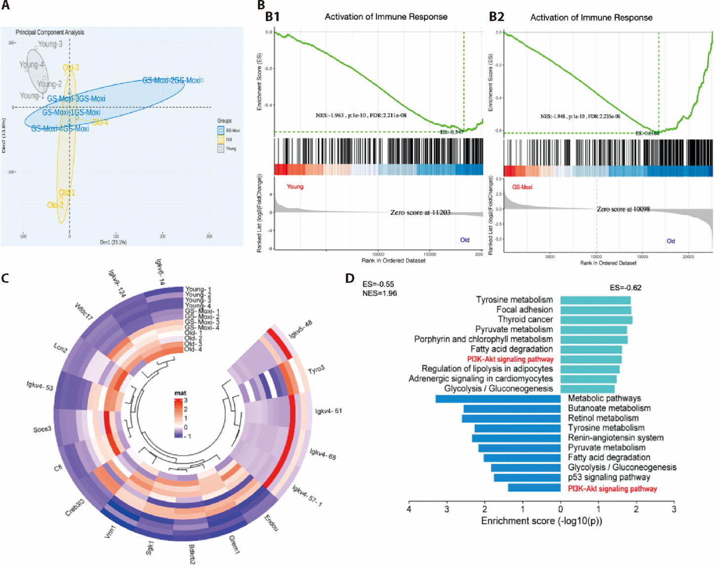
Figure 3 RNA-seq results of GS-Moxi-treated aging-induced mice A: PCA analysis; B: GSEA showed the differentially expressed gene sets involved in immune response; B1: young group; B2: GS-Moxi groups; C: heatmap of different genes related immunity reaction after GS-Moxi intervention among three groups. Red areas represent high expression, and blue areas represent low expression; D: KEGG pathway enrichment of differential gene expression among three groups. The PI3K/Akt/NF-κB signaling pathway was marked in red. Young group: untreated young mice without acupoint stimulation; Old group: naturally aged mice with spontaneous senescent renal injury; GS-Moxi group: aged mice treated with grain-sized moxibustion at Zusanli (ST36) acupoint for 12 consecutive weeks. GS-Moxi: grain-sized moxibustion; PCA: principal component analysis; GSEA: gene set enrichment analysis; KEGG: kyoto encyclopedia of genes and genomes; PI3K: phosphatidylinositol 3-kinase; AKT: protein kinase B; NF-κB: nuclear factor kappa-B. Sgk1: serum/ glucocorticoid regulated kinase 1; Creb3l3: cAMP responsive element binding protein 3-like 3; Igkv6-14: immunoglobulin kappa variable 6-14; Igkv9-124: immunoglobulin kappa variable 9-124; Wfdc17: WAP four-disulfide core domain 17; Lcn2: lipocalin 2; Igkv4-53: immunoglobulin kappa variable 4-53; Socs3: suppressor of cytokine signaling 3; Cfi: complement factor I; Vnn1: vanin 1; Bdkrb2: bradykinin receptor B2; Grem1: gremlin 1; Endou: poly(U) specific endonuclease; Igkv4-57-1: immunoglobulin kappa variable 4-57-1; Igkv4-68: immunoglobulin kappa variable 4-68; Igkv4-61: immunoglobulin kappa variable 4-61; Tyro3: TYRO3 protein tyrosine kinase; Igkv5-48: immunoglobulin kappa variable 5-48.
Figure 3 RNA-seq results of GS-Moxi-treated aging-induced mice A: PCA analysis; B: GSEA showed the differentially expressed gene sets involved in immune response; B1: young group; B2: GS-Moxi groups; C: heatmap of different genes related immunity reaction after GS-Moxi intervention among three groups. Red areas represent high expression, and blue areas represent low expression; D: KEGG pathway enrichment of differential gene expression among three groups. The PI3K/Akt/NF-κB signaling pathway was marked in red. Young group: untreated young mice without acupoint stimulation; Old group: naturally aged mice with spontaneous senescent renal injury; GS-Moxi group: aged mice treated with grain-sized moxibustion at Zusanli (ST36) acupoint for 12 consecutive weeks. GS-Moxi: grain-sized moxibustion; PCA: principal component analysis; GSEA: gene set enrichment analysis; KEGG: kyoto encyclopedia of genes and genomes; PI3K: phosphatidylinositol 3-kinase; AKT: protein kinase B; NF-κB: nuclear factor kappa-B. Sgk1: serum/ glucocorticoid regulated kinase 1; Creb3l3: cAMP responsive element binding protein 3-like 3; Igkv6-14: immunoglobulin kappa variable 6-14; Igkv9-124: immunoglobulin kappa variable 9-124; Wfdc17: WAP four-disulfide core domain 17; Lcn2: lipocalin 2; Igkv4-53: immunoglobulin kappa variable 4-53; Socs3: suppressor of cytokine signaling 3; Cfi: complement factor I; Vnn1: vanin 1; Bdkrb2: bradykinin receptor B2; Grem1: gremlin 1; Endou: poly(U) specific endonuclease; Igkv4-57-1: immunoglobulin kappa variable 4-57-1; Igkv4-68: immunoglobulin kappa variable 4-68; Igkv4-61: immunoglobulin kappa variable 4-61; Tyro3: TYRO3 protein tyrosine kinase; Igkv5-48: immunoglobulin kappa variable 5-48.
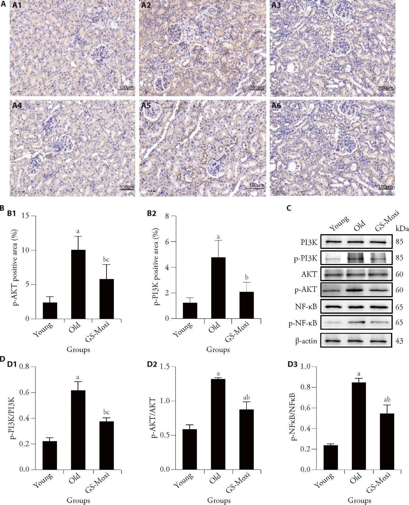
Figure 4 GS-Moxi can prevent age-related renal injury by inhibiting PI3K/Akt signaling pathway A: representative micrographs of immunohistochemical staining (scale bar, 100 µm); A1: p-PI3K in young group; A2: p-PI3K in Old group; A3: p-PI3K in Gs-Moxi group; A4: p-AKT in young group; A5: p-AKT in Old group; A6: p-AKT in Gs-Moxi group; B: immunohistochemical results in renal tissues of mice in each group; B1: p-AKT; B2: p-PI3K; C: representative Western blot bands of PI3K, p-PI3K, Akt, p-Akt, NF-κB and p-NF-κB in renal tissues. β-actin was used as a loading control; D: Western blot analysis to detect the expression levels of proteins in renal tissues; D1: p-PI3K/PI3K; D2: p-AKT/AKT; D3: p-NF-κB/NF-κB. Young group: untreated young mice without acupoint stimulation; Old group: naturally aged mice with spontaneous senescent renal injury; GS-Moxi group: aged mice treated with grain-sized moxibustion at Zusanli (ST36) acupoint for 12 consecutive weeks. GS-Moxi: grain-sized moxibustion; IHC: immunohistochemistry; PI3K: phosphatidylinositol 3-kinase; p-PI3K: phosphorylated phosphatidylinositol 3-kinase; AKT: protein kinase B; p-AKT: phosphorylated protein kinase B; NF-κB: nuclear factor kappa-B; p-NF-κB: phosphorylated nuclear factor kappa-B. Statistical analyses were measured by one-way analysis, and least significance difference test was performed for inter-group comparisons. Data were presented as mean ± standard deviation (n = 3). Compared with the young group, aP < 0.01, cP < 0.05; compared with the old group, bP < 0.01.
Figure 4 GS-Moxi can prevent age-related renal injury by inhibiting PI3K/Akt signaling pathway A: representative micrographs of immunohistochemical staining (scale bar, 100 µm); A1: p-PI3K in young group; A2: p-PI3K in Old group; A3: p-PI3K in Gs-Moxi group; A4: p-AKT in young group; A5: p-AKT in Old group; A6: p-AKT in Gs-Moxi group; B: immunohistochemical results in renal tissues of mice in each group; B1: p-AKT; B2: p-PI3K; C: representative Western blot bands of PI3K, p-PI3K, Akt, p-Akt, NF-κB and p-NF-κB in renal tissues. β-actin was used as a loading control; D: Western blot analysis to detect the expression levels of proteins in renal tissues; D1: p-PI3K/PI3K; D2: p-AKT/AKT; D3: p-NF-κB/NF-κB. Young group: untreated young mice without acupoint stimulation; Old group: naturally aged mice with spontaneous senescent renal injury; GS-Moxi group: aged mice treated with grain-sized moxibustion at Zusanli (ST36) acupoint for 12 consecutive weeks. GS-Moxi: grain-sized moxibustion; IHC: immunohistochemistry; PI3K: phosphatidylinositol 3-kinase; p-PI3K: phosphorylated phosphatidylinositol 3-kinase; AKT: protein kinase B; p-AKT: phosphorylated protein kinase B; NF-κB: nuclear factor kappa-B; p-NF-κB: phosphorylated nuclear factor kappa-B. Statistical analyses were measured by one-way analysis, and least significance difference test was performed for inter-group comparisons. Data were presented as mean ± standard deviation (n = 3). Compared with the young group, aP < 0.01, cP < 0.05; compared with the old group, bP < 0.01.
| [1] | Zelniker TA, Wiviott SD, Raz I, et al. SGLT2 inhibitors for primary and secondary prevention of cardiovascular and renal outcomes in type 2 diabetes: a systematic review and Meta-analysis of cardiovascular outcome trials. Lancet 2019; 39310166: 31-9. |
| [2] | Yang M, Liu Y, Luo SL, et al. DsbA-L ameliorates renal aging and renal fibrosis by maintaining mitochondrial homeostasis. Acta Pharmacol Sin 2024; 454: 777-89. |
| [3] | Ruiz-Ortega M, Rayego-Mateos S, Lamas S, Ortiz A, Rodrigues-Diez RR. Targeting the progression of chronic kidney disease. Nat Rev Nephrol 2020; 165: 269-88. |
| [4] | Tanriover C, Copur S, Mutlu A, et al. Early aging and premature vascular aging in chronic kidney disease. Clin Kidney J 2023; 1611: 1751-65. |
| [5] | Jankowski J, Floege J, Fliser D, et al. Cardiovascular disease in chronic kidney disease pathophysiological insights and therapeutic options. Circulation 2021; 14311: 1157-72. |
| [6] | Wang J, Wong Y-K, Liao F. What has Traditional Chinese Medicine delivered for modern medicine? Expert Rev Mol Med 2018; 20: e4. |
| [7] | Wang ZQ, Xu MW, Shi Z, et al. Mild moxibustion for irritable bowel syndrome with diarrhea (IBS-D): a randomized controlled trial. J Ethnopharmacol 2022; 289: 115064. |
| [8] | Yang XT, Wang WQ, Zhang Y, Wang JT, Huang FJ. Moxibustion improves ovary function by suppressing apoptosis events and upregulating antioxidant defenses in natural aging ovary. Life Sci 2019; 229: 166-72. |
| [9] | Ouyang X, Duan H, Jin Q, et al. Moxibustion may delay the aging process of wistar rats by regulating intestinal microbiota. Biomed Pharmacother 2022; 146: 112147. |
| [10] | Lu G, Li HX, Song ZW, et al. Combination of bone marrow mesenchymal stem cells and moxibustion restores cyclophosphamide-induced premature ovarian insufficiency by improving mitochondrial function and regulating mitophagy. Stem Cell Res Ther 2024; 151: 102. |
| [11] | Sun H, Qiu MY, Li BQ, et al. Effect of moxibustion on quality of life in hemodialysis patients. Zhong Guo Zhen Jiu 2008; 285: 321-4. |
| [12] | Li N, Qiu MY, Hao JR, et al. Randomized controlled trail on moxibustion for maintenance hemodialysis patients in deficiency syndrome. Zhong Guo Zhen Jiu 2011; 311: 15-8. |
| [13] | Zhao SY, Liu WB, Wu HG, et al. Protective effect of mild moxibustion at zusanli on kidney and effect on FN and TGF-β1 expression in naturally aging rats. Shi Jie Zhong Yi Yao 2022; 1712: 1719-24. |
| [14] | Jiao ZH, Gu Y, LI LN, et al. Liang regulation effect of moxibustion with small moxa-cone about the size of a wheat grain at “Zusanli” point on cellular immune function in elderly bedridden patients. Zhong Guo Zhong Yi Yao Ke Xue Ji Shu 2020; 271: 59-60. |
| [15] | Hu D, Shen W, Gong C, et al. Grain-sized moxibustion promotes NK cell antitumour immunity by inhibiting adrenergic signalling in non-small cell lung cancer. J Cell Mol Med 2021; 256: 2900-8. |
| [16] | Nie RR, Huang CH, Fu WB. Observation of therapeutic effect on post stroke depression treated by differentiation of spleen and stomach. Zhong Guo Zhen Jiu 2011; 314: 325-8. |
| [17] | Liu J, Liu J, Wang XS, et al. Comparative study of different dosages of grain-sized moxibustion on uterine artery blood flow in patients with cold and dampness primary dysmenorrhea. Zhen Ci Yan Jiu 2024; 497: 760-6. |
| [18] | Jiang Z, Zhou X, Li R, et al. Whole transcriptome analysis with sequencing: methods, challenges and potential solutions. Cell Mol Life Sci 2015; 7218: 3425-39. |
| [19] | Zhao W, Li J, Li Y, et al. Preventive effect of collagen peptides from on acute kidney injury through attenuation of oxidative stress and inflammation. Oxid Med Cell Longev 2022; 2022: 8186838. |
| [20] | Wang MX, Wang MM, Liu C, et al. A geniposide-phospholipid complex ameliorates posthyperuricemia chronic kidney disease induced by inflammatory reactions and oxidative stress. Eur J Pharmacol 2022; 930: 175157. |
| [21] | Li J, Zhang K, Xu M, et al. Baicalin - 2- ethoxyethyl ester alleviates renal fibrosis by inhibiting PI3K/AKT/NF-kappa B signaling pathway. Toxicol Appl Pharmacol 2024; 483: 116827. |
| [22] | LoRusso PM. Inhibition of the PI3K/AKT/mTOR pathway in solid tumors. J Clin Oncol 2016; 3431: 3803-15. |
| [23] | Song Z, Zhu M, Wu J, et al. Fucoidans from cucumaria frondosa ameliorate renal interstitial fibrosis via inhibition of the PI3K/Akt/NF-kappa B signaling pathway. Food Funct 2022; 133: 1168-79. |
| [24] | Zhang N, Li Z, Mu W, et al. Calorie restriction-induced SIRT6 activation delays aging by suppressing NF-kappa B signaling. Cell Cycle 2016; 157: 1009-18. |
| [25] | Xu L, Wang J, Yu H, et al. GLIS1 alleviates cell senescence and renal fibrosis through PGC1-alpha mediated mitochondrial quality control in kidney aging. Free Radic Biol Med 2023; 209Pt 1: 171-84. |
| [26] | Miao J, Liu J, Niu J, et al. Wnt/beta-catenin/RAS signaling mediates age-related renal fibrosis and is associated with mitochondrial dysfunction. Aging Cell 2019; 185: e13004. |
| [27] | Kim H, Yu MR, Lee H, et al. Metformin inhibits chronic kidney disease-induced DNA damage and senescence of mesenchymal stem cells. Aging Cell 2021; 202: e13317. |
| [28] | Fang Y, Gong AY, Haller ST, et al. The ageing kidney: molecular mechanisms and clinical implications. Ageing Res Rev 2020; 63: 101151. |
| [29] | Guo X, Xin Q, Wei P, et al. Antioxidant and anti-aging activities of longan crude and purified polysaccharide (LP-A) in nematode caenorhabditis elegans. Int J Biol Macromol 2024; 267Pt 2: 131634. |
| [30] | Rahman MA, Shuvo AA, Bepari AK, et al. Curcumin improves D-galactose and normal-aging associated memory impairment in mice: in vivo and in silico-based studies. PLoS One 2022; 176: e0270123. |
| [31] | Castel P, Ellis H, Bago R, et al. PDK1-SGK1 signaling sustains AKT-independent mTORC1 activation and confers resistance to PI3Kalpha inhibition. Cancer Cell 2016; 302: 229-42. |
| [32] | Dattilo V, D'Antona L, Talarico C, et al. SGK 1 affects RAN/RANBP1/RANGAP1 via SP1 to play a critical role in pre-miRNA nuclear export: a new route of epigenomic regulation. Sci Rep 2017; 7: 45361. |
| [33] | Abbruzzese C, Mattarocci S, Pizzuti L, et al. Determination of SGK1 mRNA in non-small cell lung cancer samples underlines high expression in squamous cell carcinomas. J Exp Clin Cancer Res 2012; 311: 4. |
| [34] | Spagnuolo R, Dattilo V, D'Antona L, et al. Deregulation of SGK1 in ulcerative colitis: a paradoxical relationship between immune cells and colonic epithelial cells. Inflamm Bowel Dis 2018; 249: 1967-77. |
| [35] | Lang F, Voelkl J. Therapeutic potential of serum and glucocorticoid inducible kinase inhibition. Expert Opin Investig Drugs 2013; 226: 701-14. |
| [36] | Zhuang L, Jin G, Hu X, et al. The inhibition of SGK1 suppresses epithelial-mesenchymal transition and promotes renal tubular epithelial cell autophagy in diabetic nephropathy. Am J Transl Res 2019; 118: 4946-56. |
| [37] | Wang J, Zhu H, Huang L, et al. Nrf2 signaling attenuates epithelial-to-mesenchymal transition and renal interstitial fibrosis via PI3K/Akt signaling pathways. Exp Mol Pathol 2019; 111: 104296. |
| [38] | Wang Z, Chen Z, Li B, et al. Curcumin attenuates renal interstitial fibrosis of obstructive nephropathy by suppressing epithelial-mesenchymal transition through inhibition of the TLR4/NF-small ka, CyrillicB and PI3K/AKT signalling pathways. Pharm Biol 2020; 581: 828-37. |
| [39] | Li D, Yu K, Feng F, et al. Hydroxychloroquine alleviates renal interstitial fibrosis by inhibiting the PI3K/Akt signaling pathway. Biochem Biophys Res Commun 2022; 610: 154-61. |
| [40] | Jung KJ, Min KJ, Park JW, et al. Carnosic acid attenuates unilateral ureteral obstruction-induced kidney fibrosis via inhibition of Akt-mediated Nox4 expression. Free Radic Biol Med 2016; 97: 50-7. |
| [41] | Dou F, Liu Y, Liu L, et al. Aloe-Emodin ameliorates renal fibrosis via inhibiting PI3K/Akt/mTOR signaling pathway in vivo and in vitro. Rejuvenation Res 2019; 223: 218-29. |
| [42] | Zhu JF, Huang W, Yi HM, et al. Annexin A1-suppressed autophagy promotes nasopharyngeal carcinoma cell invasion and metastasis by PI3K/AKT signaling activation. Cell Death Dis 2018; 912: 1154. |
| [43] | Liang F, Ren C, Wang J, et al. The crosstalk between STAT3 and p53/RAS signaling controls cancer cell metastasis and cisplatin resistance via the Slug/MAPK/PI3K/AKT-mediated regulation of EMT and autophagy. Oncogenesis 2019; 810: 59. |
| [44] | Xu WT, Yang Z, Lu NH. A new role for the PI3K/Akt signaling pathway in the epithelial-mesenchymal transition. Cell Adh Migr 2015; 94: 317-24. |
| [45] | White S, Lin L, Hu KB. NF-kappaB and tPA Signaling in kidney and other diseases. Cells 2020; 9: 1348. |
| Viewed | ||||||
|
Full text |
|
|||||
|
Abstract |
|
|||||