Journal of Traditional Chinese Medicine ›› 2023, Vol. 43 ›› Issue (3): 457-465.DOI: 10.19852/j.cnki.jtcm.20220707.005
Previous Articles Next Articles
Salvianolic acid B promotes the invasion and migration of H2O2-induced HTR-8/Svneo trophoblast cells by upregulating matrix metalloproteinase-9 via the phosphatidylinositol-4,5-bisphosphate 3-kinase/protein kinase B pathway
ZHAO Zhiqiang, ZHANG Chong, ZHU Yunxia( )
)
- Department of Gynecology and Obstetrics, Beijing You'an Hospital of Capital Medical University, Beijing 100069, China
-
Received:2021-06-27Accepted:2021-09-30Online:2023-06-15Published:2022-07-07 -
Contact:ZHU Yunxia, Beijing You'an Hospital of Capital Medical University, Beijing 100069, China. yunxiadyxh@163.com. Telephone: +86-13522674990
Cite this article
ZHAO Zhiqiang, ZHANG Chong, ZHU Yunxia. Salvianolic acid B promotes the invasion and migration of H2O2-induced HTR-8/Svneo trophoblast cells by upregulating matrix metalloproteinase-9 via the phosphatidylinositol-4,5-bisphosphate 3-kinase/protein kinase B pathway[J]. Journal of Traditional Chinese Medicine, 2023, 43(3): 457-465.
share this article
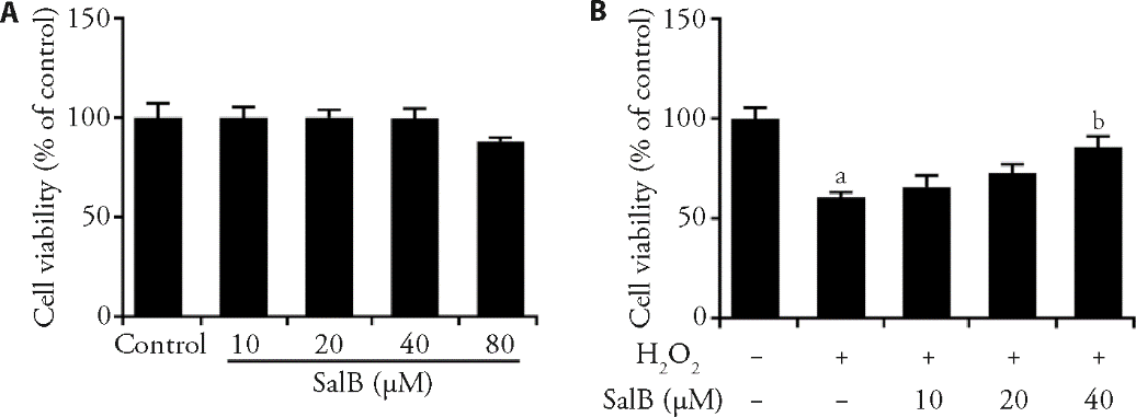
Figure 1 SalB promoted the proliferation of HTR-8/ Svneo cells A: MTT detected the cell viability after treatment with different concentrations (10, 20, 40 and 80 μM) of SalB for 24 h; B: CCK-8 detected the viability in untreated cells or in cells exposed to 300?μmol/L of H2O2 for 3 h alone or in cells treated by different concentrations (10, 20 and 40 μM) of SalB for 24 h after being exposed to 300?μmol/L of H2O2 for 3 h. aP<0.001 vs Control; bP<0.001 vs H2O2. SalB: salvianolic acid B; MTT: 3-(4,5-dimethylthiazol-2-yl)-2,5-diphenyltetrazolium bromide.
Figure 1 SalB promoted the proliferation of HTR-8/ Svneo cells A: MTT detected the cell viability after treatment with different concentrations (10, 20, 40 and 80 μM) of SalB for 24 h; B: CCK-8 detected the viability in untreated cells or in cells exposed to 300?μmol/L of H2O2 for 3 h alone or in cells treated by different concentrations (10, 20 and 40 μM) of SalB for 24 h after being exposed to 300?μmol/L of H2O2 for 3 h. aP<0.001 vs Control; bP<0.001 vs H2O2. SalB: salvianolic acid B; MTT: 3-(4,5-dimethylthiazol-2-yl)-2,5-diphenyltetrazolium bromide.

Figure 2 SalB inhibited oxidative damage and apoptosis in HTR-8/Svneo cells induced by H2O2 A. ELISA assay detected the expression of SOD, GSH-Px and MDA in untreated cells or in cells exposed to 300?μmol/L of H2O2 for 3 h alone or in cells treated by different concentrations (10, 20 and 40 μM) of SalB for 24 h after being exposed to 300?μmol/L of H2O2 for 3 h. B. TUNEL assay detected the apoptosis in untreated cells or in cells exposed to 300?μmol/L of H2O2 for 3 h alone or in cells treated by different concentrations (10, 20 and 40 μM) of SalB for 24 h after being exposed to 300?μmol/L of H2O2 for 3 h. magnification, x200. C. Western blot detected the expression of apoptosis-related proteins in untreated cells or in cells exposed to 300?μmol/L of H2O2 for 3 h alone or in cells treated by different concentrations (10, 20 and 40 μM) of SalB for 24 h after being exposed to 300?μmol/L of H2O2 for 3 h. aP<0.001 vs control; bP<0.05, dP<0.01, cP<0.001 vs H2O2. SalB: salvianolic acid B; SOD: superoxide dismutase; GSH-Px: glutathione-Px; MDA: malondialdehyde; Bcl-2: B cell lymphoma-2; Bax: Bcl-2 associated X; GAPDH: glyceraldehyde-3-phosphate dehydrogenase; ELISA: enzyme-linked immunosorbent assay; TUNEL: terminal deoxynucleotidyl transferase (TdT) dUTP nick-end labeling; DAPI: 4',6-diamidino-2-phenylindole.
Figure 2 SalB inhibited oxidative damage and apoptosis in HTR-8/Svneo cells induced by H2O2 A. ELISA assay detected the expression of SOD, GSH-Px and MDA in untreated cells or in cells exposed to 300?μmol/L of H2O2 for 3 h alone or in cells treated by different concentrations (10, 20 and 40 μM) of SalB for 24 h after being exposed to 300?μmol/L of H2O2 for 3 h. B. TUNEL assay detected the apoptosis in untreated cells or in cells exposed to 300?μmol/L of H2O2 for 3 h alone or in cells treated by different concentrations (10, 20 and 40 μM) of SalB for 24 h after being exposed to 300?μmol/L of H2O2 for 3 h. magnification, x200. C. Western blot detected the expression of apoptosis-related proteins in untreated cells or in cells exposed to 300?μmol/L of H2O2 for 3 h alone or in cells treated by different concentrations (10, 20 and 40 μM) of SalB for 24 h after being exposed to 300?μmol/L of H2O2 for 3 h. aP<0.001 vs control; bP<0.05, dP<0.01, cP<0.001 vs H2O2. SalB: salvianolic acid B; SOD: superoxide dismutase; GSH-Px: glutathione-Px; MDA: malondialdehyde; Bcl-2: B cell lymphoma-2; Bax: Bcl-2 associated X; GAPDH: glyceraldehyde-3-phosphate dehydrogenase; ELISA: enzyme-linked immunosorbent assay; TUNEL: terminal deoxynucleotidyl transferase (TdT) dUTP nick-end labeling; DAPI: 4',6-diamidino-2-phenylindole.
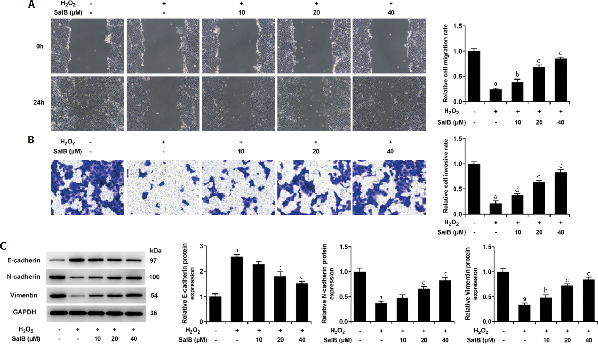
Figure 3 SalB promoted invasion, migration and EMT of HTR-8/ Svneo cell induced by H2O2. A. Wound healing detected the cell migration ability in untreated cells or in cells exposed to 300?μmol/L of H2O2 for 3 h alone or in cells treated by different concentrations (10, 20 and 40 μM) of SalB for 24 h after being exposed to 300?μmol/L of H2O2 for 3 h. magnification, x100. B. Transwell detected the cell invasion ability in untreated cells or in cells exposed to 300?μmol/L of H2O2 for 3 h alone or in cells treated by different concentrations (10, 20 and 40 μM) of SalB for 24 h after being exposed to 300?μmol/L of H2O2 for 3 h. magnification, x100. C. Western blot detected the expression of EMT-related proteins in untreated cells or in cells exposed to 300?μmol/L of H2O2 for 3 h alone or in cells treated by different concentrations (10, 20 and 40 μM) of SalB for 24 h after being exposed to 300?μmol/L of H2O2 for 3 h. aP<0.001 vs Control; bP<0.05, dP<0.01, cP<0.001 vs H2O2. SalB: salvianolic acid B; GAPDH: glyceraldehyde-3-phosphate dehydrogenase.
Figure 3 SalB promoted invasion, migration and EMT of HTR-8/ Svneo cell induced by H2O2. A. Wound healing detected the cell migration ability in untreated cells or in cells exposed to 300?μmol/L of H2O2 for 3 h alone or in cells treated by different concentrations (10, 20 and 40 μM) of SalB for 24 h after being exposed to 300?μmol/L of H2O2 for 3 h. magnification, x100. B. Transwell detected the cell invasion ability in untreated cells or in cells exposed to 300?μmol/L of H2O2 for 3 h alone or in cells treated by different concentrations (10, 20 and 40 μM) of SalB for 24 h after being exposed to 300?μmol/L of H2O2 for 3 h. magnification, x100. C. Western blot detected the expression of EMT-related proteins in untreated cells or in cells exposed to 300?μmol/L of H2O2 for 3 h alone or in cells treated by different concentrations (10, 20 and 40 μM) of SalB for 24 h after being exposed to 300?μmol/L of H2O2 for 3 h. aP<0.001 vs Control; bP<0.05, dP<0.01, cP<0.001 vs H2O2. SalB: salvianolic acid B; GAPDH: glyceraldehyde-3-phosphate dehydrogenase.
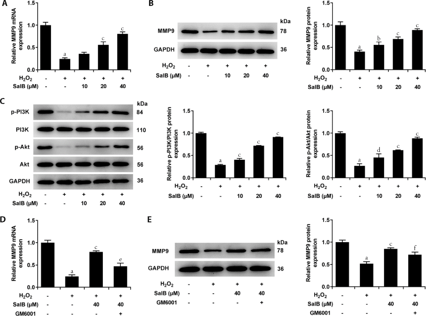
Figure 4 SalB upregulated the expression of MMP-9 in H2O2-induced HTR-8/Svneo cells through PI3K/Akt pathway. A. RT-qPCR detected the expression of MMP9 in untreated cells or in cells exposed to 300?μmol/L of H2O2 for 3 h alone or in cells treated by different concentrations (10, 20 and 40 μM) of SalB for 24 h after being exposed to 300?μmol/L of H2O2 for 3 h. B. Western blot detected the expression of MMP9 in untreated cells or in cells exposed to 300?μmol/L of H2O2 for 3 h alone or in cells treated by different concentrations (10, 20 and 40 μM) of SalB for 24 h after being exposed to 300?μmol/L of H2O2 for 3 h. C. Western blot detected the expression of PI3K/AKT in untreated cells or in cells exposed to 300?μmol/L of H2O2 for 3 h alone or in cells treated by different concentrations (10, 20 and 40 μM) of SalB for 24 h after being exposed to 300?μmol/L of H2O2 for 3 h. D. RT-qPCR detected the expression of MMP9 in untreated cells or in cells exposed to 300?μmol/L of H2O2 for 3 h alone or in cells treated by 40 μM of SalB for 24 h after being exposed to 300?μmol/L of H2O2 for 3 h or in cells co-treated by SalB (40 μM) and LY294002 for 24 h after being exposed to 300?μmol/L of H2O2 for 3 h. E. Western blot detected the expression of MMP9 in untreated cells or in cells exposed to 300?μmol/L of H2O2 for 3 h alone or in cells treated by 40 μM of SalB for 24 h after being exposed to 300?μmol/L of H2O2 for 3 h or in cells co-treated by SalB (40 μM) and LY294002 for 24 h after being exposed to 300?μmol/L of H2O2 for 3 h. aP<0.001 vs Control; dP<0.01, cP<0.001 vs H2O2; fP<0.05, eP<0.001 vs H2O2 + SalB. SalB: salvianolic acid B; MMP9, Matrix metallopeptidase 9; p-PI3K: phosphorylated phosphatidylinositol-3-kinase; PI3K: phosphatidylinositol-4,5-bisphosphate 3-kinase; p-Akt: phosphorylated protein kinase B; Akt: protein kinase B; GAPDH: glyceraldehyde-3-phosphate dehydrogenase; RT-qPCR: reverse transcription-quantitative real-time polymerase chain reaction.
Figure 4 SalB upregulated the expression of MMP-9 in H2O2-induced HTR-8/Svneo cells through PI3K/Akt pathway. A. RT-qPCR detected the expression of MMP9 in untreated cells or in cells exposed to 300?μmol/L of H2O2 for 3 h alone or in cells treated by different concentrations (10, 20 and 40 μM) of SalB for 24 h after being exposed to 300?μmol/L of H2O2 for 3 h. B. Western blot detected the expression of MMP9 in untreated cells or in cells exposed to 300?μmol/L of H2O2 for 3 h alone or in cells treated by different concentrations (10, 20 and 40 μM) of SalB for 24 h after being exposed to 300?μmol/L of H2O2 for 3 h. C. Western blot detected the expression of PI3K/AKT in untreated cells or in cells exposed to 300?μmol/L of H2O2 for 3 h alone or in cells treated by different concentrations (10, 20 and 40 μM) of SalB for 24 h after being exposed to 300?μmol/L of H2O2 for 3 h. D. RT-qPCR detected the expression of MMP9 in untreated cells or in cells exposed to 300?μmol/L of H2O2 for 3 h alone or in cells treated by 40 μM of SalB for 24 h after being exposed to 300?μmol/L of H2O2 for 3 h or in cells co-treated by SalB (40 μM) and LY294002 for 24 h after being exposed to 300?μmol/L of H2O2 for 3 h. E. Western blot detected the expression of MMP9 in untreated cells or in cells exposed to 300?μmol/L of H2O2 for 3 h alone or in cells treated by 40 μM of SalB for 24 h after being exposed to 300?μmol/L of H2O2 for 3 h or in cells co-treated by SalB (40 μM) and LY294002 for 24 h after being exposed to 300?μmol/L of H2O2 for 3 h. aP<0.001 vs Control; dP<0.01, cP<0.001 vs H2O2; fP<0.05, eP<0.001 vs H2O2 + SalB. SalB: salvianolic acid B; MMP9, Matrix metallopeptidase 9; p-PI3K: phosphorylated phosphatidylinositol-3-kinase; PI3K: phosphatidylinositol-4,5-bisphosphate 3-kinase; p-Akt: phosphorylated protein kinase B; Akt: protein kinase B; GAPDH: glyceraldehyde-3-phosphate dehydrogenase; RT-qPCR: reverse transcription-quantitative real-time polymerase chain reaction.

Figure 5 MMP-9 inhibitor GM6001 reversed the oxidative damage and apoptosis of SalB on HTR-8/ Svneo cells induced by H2O2 A: ELISA assay detected the expression of SOD, GSH-Px and MDA in untreated cells or in cells exposed to 300?μmol/L of H2O2 for 3 h alone or in cells treated by 40 μM of SalB for 24 h after being exposed to 300?μmol/L of H2O2 for 3 h or in cells co-treated by SalB (40 μM) and GM6001 for 24 h after being exposed to 300?μmol/L of H2O2 for 3 h; B:TUNEL assay detected the apoptosis in untreated cells or in cells exposed to 300?μmol/L of H2O2 for 3 h alone or in cells treated by 40 μM of SalB for 24 h after being exposed to 300?μmol/L of H2O2 for 3 h or in cells co-treated by SalB (40 μM) and GM6001 for 24 h after being exposed to 300?μmol/L of H2O2 for 3 h. magnification, ×200; C: Western blot detected the expression of apoptosis-related proteins in untreated cells or in cells exposed to 300?μmol/L of H2O2 for 3 h alone or in cells treated by 40 μM of SalB for 24 h after being exposed to 300?μmol/L of H2O2 for 3 h or in cells co-treated by SalB (40 μM) and GM6001 for 24 h after being exposed to 300?μmol/L of H2O2 for 3 h. aP<0.001 vs Control; bP<0.001 vs H2O2; cP<0.05, dP<0.01, eP<0.001 vs H2O2 + SalB. SalB: salvianolic acid B; SOD: superoxide dismutase; GSH-Px: glutathione-Px; MDA: malondialdehyde; Bcl-2: B cell lymphoma-2; Bax: Bcl-2 associated X; GAPDH: glyceraldehyde-3-phosphate dehydrogenase; ELISA: enzyme-linked immunosorbent assay; TUNEL: terminal deoxynucleotidyl transferase (TdT) dUTP nick-end labeling; Dapi: 4',6-diamidino-2-phenylindole.
Figure 5 MMP-9 inhibitor GM6001 reversed the oxidative damage and apoptosis of SalB on HTR-8/ Svneo cells induced by H2O2 A: ELISA assay detected the expression of SOD, GSH-Px and MDA in untreated cells or in cells exposed to 300?μmol/L of H2O2 for 3 h alone or in cells treated by 40 μM of SalB for 24 h after being exposed to 300?μmol/L of H2O2 for 3 h or in cells co-treated by SalB (40 μM) and GM6001 for 24 h after being exposed to 300?μmol/L of H2O2 for 3 h; B:TUNEL assay detected the apoptosis in untreated cells or in cells exposed to 300?μmol/L of H2O2 for 3 h alone or in cells treated by 40 μM of SalB for 24 h after being exposed to 300?μmol/L of H2O2 for 3 h or in cells co-treated by SalB (40 μM) and GM6001 for 24 h after being exposed to 300?μmol/L of H2O2 for 3 h. magnification, ×200; C: Western blot detected the expression of apoptosis-related proteins in untreated cells or in cells exposed to 300?μmol/L of H2O2 for 3 h alone or in cells treated by 40 μM of SalB for 24 h after being exposed to 300?μmol/L of H2O2 for 3 h or in cells co-treated by SalB (40 μM) and GM6001 for 24 h after being exposed to 300?μmol/L of H2O2 for 3 h. aP<0.001 vs Control; bP<0.001 vs H2O2; cP<0.05, dP<0.01, eP<0.001 vs H2O2 + SalB. SalB: salvianolic acid B; SOD: superoxide dismutase; GSH-Px: glutathione-Px; MDA: malondialdehyde; Bcl-2: B cell lymphoma-2; Bax: Bcl-2 associated X; GAPDH: glyceraldehyde-3-phosphate dehydrogenase; ELISA: enzyme-linked immunosorbent assay; TUNEL: terminal deoxynucleotidyl transferase (TdT) dUTP nick-end labeling; Dapi: 4',6-diamidino-2-phenylindole.

Figure 6 MMP-9 inhibitor GM6001 reversed the invasion, migration and EMT of SalB on HTR-8/Svneo cells induced by H2O2 A: wound healing detected the cell migration ability in untreated cells or in cells exposed to 300?μmol/L of H2O2 for 3 h alone or in cells treated by 40 μM of SalB for 24 h after being exposed to 300?μmol/L of H2O2 for 3 h or in cells co-treated by SalB (40 μM) and GM6001 for 24 h after being exposed to 300?μmol/L of H2O2 for 3 h. magnification, ×100; B: transwell detected the cell invasion ability in untreated cells or in cells exposed to 300?μmol/L of H2O2 for 3 h alone or in cells treated by 40 μM of SalB for 24 h after being exposed to 300?μmol/L of H2O2 for 3 h or in cells co-treated by SalB (40 μM) and GM6001 for 24 h after being exposed to 300?μmol/L of H2O2 for 3 h. magnification, ×100; C: Western blot detected the expression of EMT-related proteins in untreated cells or in cells exposed to 300?μmol/L of H2O2 for 3 h alone or in cells treated by 40 μM of SalB for 24 h after being exposed to 300?μmol/L of H2O2 for 3 h or in cells co-treated by SalB (40 μM) and GM6001 for 24 h after being exposed to 300?μmol/L of H2O2 for 3 h. aP<0.001 vs control; bP<0.001 vs H2O2; eP<0.05, dP<0.01, cP<0.001 vs H2O2 + SalB. SalB: salvianolic acid B; GAPDH: glyceraldehyde-3-phosphate dehydrogenase.
Figure 6 MMP-9 inhibitor GM6001 reversed the invasion, migration and EMT of SalB on HTR-8/Svneo cells induced by H2O2 A: wound healing detected the cell migration ability in untreated cells or in cells exposed to 300?μmol/L of H2O2 for 3 h alone or in cells treated by 40 μM of SalB for 24 h after being exposed to 300?μmol/L of H2O2 for 3 h or in cells co-treated by SalB (40 μM) and GM6001 for 24 h after being exposed to 300?μmol/L of H2O2 for 3 h. magnification, ×100; B: transwell detected the cell invasion ability in untreated cells or in cells exposed to 300?μmol/L of H2O2 for 3 h alone or in cells treated by 40 μM of SalB for 24 h after being exposed to 300?μmol/L of H2O2 for 3 h or in cells co-treated by SalB (40 μM) and GM6001 for 24 h after being exposed to 300?μmol/L of H2O2 for 3 h. magnification, ×100; C: Western blot detected the expression of EMT-related proteins in untreated cells or in cells exposed to 300?μmol/L of H2O2 for 3 h alone or in cells treated by 40 μM of SalB for 24 h after being exposed to 300?μmol/L of H2O2 for 3 h or in cells co-treated by SalB (40 μM) and GM6001 for 24 h after being exposed to 300?μmol/L of H2O2 for 3 h. aP<0.001 vs control; bP<0.001 vs H2O2; eP<0.05, dP<0.01, cP<0.001 vs H2O2 + SalB. SalB: salvianolic acid B; GAPDH: glyceraldehyde-3-phosphate dehydrogenase.
| [1] |
Filipek A, Jurewicz E. Preeclampsia-a disease of pregnant women. Postepy Biochem 2018; 64: 232-29.
DOI PMID |
| [2] |
Ramos JGL, Sass N, Costa SHM. Preeclampsia. Rev Bras Ginecol Obstet 2017; 39: 496-512.
DOI URL |
| [3] |
Guedes-Martins L. Superimposed preeclampsia. Adv Exp Med Biol 2017; 956: 409-17.
DOI PMID |
| [4] |
Jiang P, Guo Y, Dang R, et al. Salvianolic acid B protects against lipopolysaccharide-induced behavioral deficits and neuroinflammatory response: involvement of autophagy and NLRP3 inflammasome. J Neuroinflammation 2017; 14: 239.
DOI URL |
| [5] |
Liu Q, Lu J, Lin J, et al. Salvianolic acid B attenuates experimental skin fibrosis of systemic sclerosis. Biomed Pharmacother 2019; 110: 546-53.
DOI PMID |
| [6] | Li K, Song J, Zhao Q, et al. Effective component of Salvia miltiorrhiza in promoting cardiomyogenic differentiation of human placentaderived mesenchymal stem cells. Int J Mol Med 2018; 41: 962-8. |
| [7] | Wang J, Liu C, Que W, et al. Immunomodulatory effects of Salvianolic acid B in a spontaneous abortion mouse model. J Reprod Immunol 2020; 137: 103075. |
| [8] | Suo M, Sun Y, Yang H, et al. MiR-183-5p suppressed the invasion and migration of HTR-8/SVneo trophoblast cells partly via targeting MMP-9 in preeclampsia. Biosci Rep 2020; 40: BSR20192575. |
| [9] |
Huang W, Lu W, Li Q, et al. Effects of cyclosporine A on proliferation, invasion and migration of HTR-8/SVneo human extravillous trophoblasts. Biochem Biophys Res Commun 2020; 533: 645-50.
DOI URL |
| [10] |
Daina A, Michielin O, Zoete V. SwissTargetPrediction: updated data and new features for efficient prediction of protein targets of small molecules. Nucleic Acids Res 2019; 47: W357-64.
DOI URL |
| [11] |
Li T, Wei S, Fan C, Tang D, Luo D. Nesfatin-1 promotes proliferation, migration and invasion of HTR-8/SVneo trophoblast cells and inhibits oxidative stress via activation of PI3K/AKT/mTOR and AKT/GSK3beta pathway. Reprod Sci 2021; 28: 550-61.
DOI |
| [12] |
Yang C, Luo L, Bai X, et al. Highly-expressed micoRNA-21 in adipose derived stem cell exosomes can enhance the migration and proliferation of the HaCaT cells by increasing the MMP-9 expression through the PI3K/AKT pathway. Arch Biochem Biophys 2020; 681: 108259.
DOI URL |
| [13] | Li M, Sun T, Wu X, An P, Wu X, Dang H. Autophagy in the HTR-8/SVneo cell oxidative stress model is associated with the NLRP1 inflammasome. Oxid Med Cell Longev 2021; 2021: 2353504. |
| [14] |
Guo H, Wang Y, Liu D. Silibinin ameliorats H2O2-induced cell apoptosis and oxidative stress response by activating Nrf2 signaling in trophoblast cells. Acta Histochem 2020; 122: 151620.
DOI URL |
| [15] |
Abbas Y, Turco MY, Burton GJ, Moffett A. Investigation of human trophoblast invasion in vitro. Hum Reprod Update 2020; 26: 501-13.
DOI URL |
| [16] |
Shi J, Guo S, Wu Y, Chen G, Lai J, Xu X. Behaviour of cell penetrating peptide TAT-modified liposomes loaded with salvianolic acid B on the migration, proliferation, and survival of human skin fibroblasts. J Liposome Res 2020; 30: 93-106.
DOI PMID |
| [17] |
Wang J, Ma Y, Guo M, Yang H, Guan X. Salvianolic acid B suppresses EMT and apoptosis to lessen drug resistance through AKT/mTOR in gastric cancer cells. Cytotechnology 2021; 73: 49-61.
DOI PMID |
| [18] |
Guo G, Li B, Wang Y, et al. Effects of salvianolic acid B on proliferation, neurite outgrowth and differentiation of neural stem cells derived from the cerebral cortex of embryonic mice. Sci China Life Sci 2010; 53: 653-62.
DOI PMID |
| [19] |
Espino YSS, Flores-Pliego A, Espejel-Nunez A, et al. New insights into the role of matrix metalloproteinases in preeclampsia. Int J Mol Sci 2017; 18: 1448.
DOI URL |
| [20] | Wu L, Zhao KQ, Wang W, et al. Nuclear receptor coactivator 6 promotes HTR-8/SVneo cell invasion and migration by activating NF-kappaB-mediated MMP9 transcription. Cell Prolif 2020; 53: e12876. |
| [21] |
Zhu W, Wu X, Yang B, et al. miR-188-5p regulates proliferation and invasion via PI3K/Akt/MMP-2/9 signaling in keloids. Acta Biochim Biophys Sin (Shanghai) 2019; 51: 185-96.
DOI URL |
| [22] | Yuan J, Xu XJ, Lin Y, et al. LncRNA MALAT1 expression inhibition suppresses tongue squamous cell carcinoma proliferation, migration and invasion by inactivating PI3K/Akt pathway and downregulating MMP-9 expression. Eur Rev Med Pharmacol Sci 2019; 23: 198-206. |
| Viewed | ||||||
|
Full text |
|
|||||
|
Abstract |
|
|||||