Journal of Traditional Chinese Medicine ›› 2026, Vol. 46 ›› Issue (3): 641-651.DOI: 10.19852/j.cnki.jtcm.2026.03.008
• Original Articles • Previous Articles Next Articles
Network pharmacology and experimental validation of asiaticoside protecting against diabetic kidney disease by regulating 11β-hydroxysteroid dehydrogenase type 2
ZHU Qin1, BI Peng1, ZENG Jiali1, ZHANG Yang1, HU Lidan2, CAO Zhongkai2, ZHU Jingyu1, JIN Qinyang3( )
)
- 1
Department of Nephrology (Key Laboratory of Zhejiang Province ,Management of Kidney Disease), Hangzhou Traditional Chinese Medicine Hospital Affiliated to Zhejiang Chinese Medical University Hangzhou 310007, China
2the Children’s Hospital ,Zhejiang University School of Medicine, National Clinical Research Center for Child Health Hangzhou 310052, China
3Geriatric Medicine Center ,Department of Geriatric Medicine, Zhejiang Provincial People's Hospital (Affiliated People's Hospital), Hangzhou Medical College Hangzhou 310014, China
-
Received:2024-12-22Accepted:2025-07-05Online:2026-06-15Published:2026-06-08 -
Contact:JIN Qinyang, Geriatric Medicine Center, Department of Geriatric Medicine, Zhejiang Provincial People's Hospital (Affiliated People's Hospital), Hangzhou Medical College, Hangzhou 310014, China. jqy119@163.com, Telephone: +86-13777877613 -
About author:First author contact:ZHU Qin and BI Peng are co-first authors and contributed equally to this work
-
Supported by:National Natural Science Foundation of China for Young Scholars: Study on the Mechanism of Compound Centella Asiatica Mediate 24-dehydrocholesterol Reductase (DHCR24)/Liver X Receptor (LXR) Signaling Axis to Regulate Macrophage Activation and Alleviate Microinflammation in Diabetic Kidney Disease(82205008);Medical Scientific Research Foundation of Zhejiang Province, China: Mechanistic Study of Asiaticoside in Regulating Macrophage Innate Immune Response in Diabetic Kidney Disease by Maintaining Cholesterol Homeostasis via the DHCR24/Desmosterol/ LXR Signaling Axis(2023RC242);Zhejiang Traditional Medicine and Technology Program, China: Research on the Construction of a Knowledge Graph for the Academic Thoughts and Clinical Experience of Famous Traditional Chinese Medicine Practitioner Chen Hongyu in Diagnosing and Treating Diabetic Kidney Disease(2023ZF137);Hangzhou Science and Technology Bureau project: Precise Diagnosis of Prostate Malignancies Using Artificial Intelligence(20231203A12);Special key research project of the Affiliated Hospital of Zhejiang University of Traditional Chinese Medicine: Study on Clinical Auxiliary Decision-Making Model for Professor Wang Yongjun in Diagnosis and Treatment of Diabetic Kidney Disease Based on Graph Convolutional Neural Network(2022FSYYZZ14)
Cite this article
ZHU Qin, BI Peng, ZENG Jiali, ZHANG Yang, HU Lidan, CAO Zhongkai, ZHU Jingyu, JIN Qinyang. Network pharmacology and experimental validation of asiaticoside protecting against diabetic kidney disease by regulating 11β-hydroxysteroid dehydrogenase type 2[J]. Journal of Traditional Chinese Medicine, 2026, 46(3): 641-651.
share this article
Table 1 Common targets of GO biologic processes
| Term ID | Term description | Matching proteins in network (labels) | FDR |
|---|---|---|---|
| GO:0048545 | Response to steroid hormone | NR3C1, CASP3, HSD11B2, ANXA1, ATP1A3, ATP1A1 | 3.32E-05 |
| GO:0033993 | Response to lipid | NR3C1, CASP3, HSD11B2, NFKB2, ANXA1, ATP1A3, ATP1A1 | 9.37E-05 |
| GO:0051384 | Response to glucocorticoid | NR3C1, CASP3, HSD11B2, ANXA1 | 0.0024 |
| GO:0071383 | Cellular response to steroid hormone stimulus | NR3C1, ANXA1, ATP1A3, ATP1A1 | 0.0024 |
| GO:1901700 | Response to oxygen-containing compound | NR3C1, CASP3, HSD11B2, NFKB2, ANXA1, ATP1A3, ATP1A1 | 0.0024 |
| GO:0071407 | Cellular response to organic cyclic compound | NR3C1, CASP3, ANXA1, ATP1A3, ATP1A1 | 0.0036 |
| GO:1903416 | Response to glycoside | ATP1A3, ATP1A1 | 0.0069 |
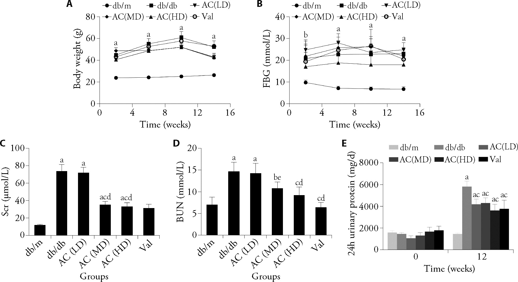
Figure 1 High-dose AC intervention alleviates the deterioration of biochemical parameters induced by DKD A: body weight; B: FBG; C: Scr; D: BUN; E: 24-h urinary protein in the 6 groups. Mice were randomly divided into six groups (n = 5 per group) and treated via oral gavage once daily for 12 weeks: control group (db/m), model group (db/db), low-dose AC group (5 g/kg), medium-dose AC group (15 g/kg), high-dose AC group (45 g/kg), and positive control group (valsartan, 5 mg/kg). All compounds were prepared as 0.2 mL suspensions for daily oral administration. AC: asiaticoside; DKD: diabetic kidney disease; FBG: fasting blood glucose; Scr: serum creatinine; BUN: blood urea nitrogen; LD: low-dose; MD: medium-dose; HD: high-dose. Differences among groups were assessed using one-way analysis of variance and the Bonferroni test. Data are presented as mean ± standard deviation (n = 5). aP < 0.01, bP < 0.05, compared with the db/m group; cP < 0.01, eP < 0.05, compared with the db/db group; dP < 0.01 compared with the AC (LD) group.
Figure 1 High-dose AC intervention alleviates the deterioration of biochemical parameters induced by DKD A: body weight; B: FBG; C: Scr; D: BUN; E: 24-h urinary protein in the 6 groups. Mice were randomly divided into six groups (n = 5 per group) and treated via oral gavage once daily for 12 weeks: control group (db/m), model group (db/db), low-dose AC group (5 g/kg), medium-dose AC group (15 g/kg), high-dose AC group (45 g/kg), and positive control group (valsartan, 5 mg/kg). All compounds were prepared as 0.2 mL suspensions for daily oral administration. AC: asiaticoside; DKD: diabetic kidney disease; FBG: fasting blood glucose; Scr: serum creatinine; BUN: blood urea nitrogen; LD: low-dose; MD: medium-dose; HD: high-dose. Differences among groups were assessed using one-way analysis of variance and the Bonferroni test. Data are presented as mean ± standard deviation (n = 5). aP < 0.01, bP < 0.05, compared with the db/m group; cP < 0.01, eP < 0.05, compared with the db/db group; dP < 0.01 compared with the AC (LD) group.
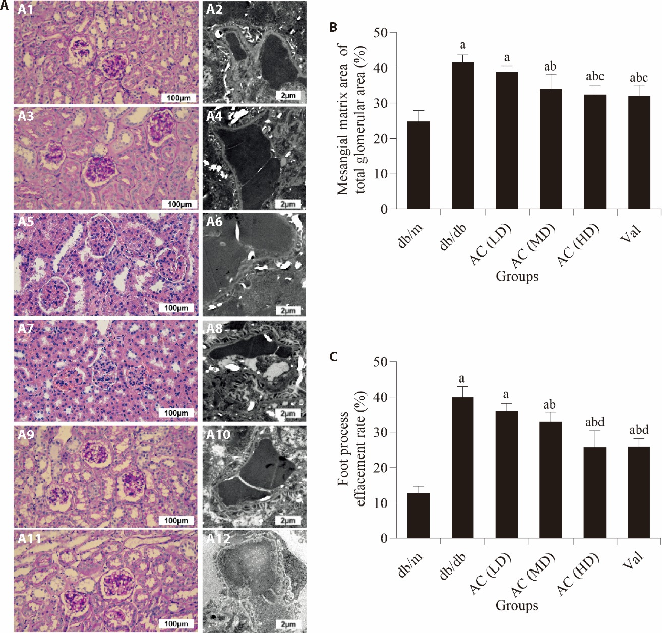
Figure 2 High-dose AC intervention ameliorated the abnormal pathological changes caused by DKD A: PAS staining and TEM; A1: db/m group; A2: db/m group; A3: db/db group; A4: db/db group; A5: AC (LD) group; A6: AC (LD) group; A7: AC (MD) group; A8: AC (MD) group; A9: AC (HD) group; A10: AC (HD) group; A11: Val group; A12: Val group; B: mesangial matrix areas; C: foot process effacement rate. Mice were randomly divided into six groups and treated via oral gavage once daily for 12 weeks: control group (db/m), model group (db/db), low-dose AC group (5 g/kg), medium-dose AC group (15 g/kg), high-dose AC group (45 g/kg), and positive control group (valsartan, 5 mg/kg). All compounds were prepared as 0.2 mL suspensions for daily oral administration. AC: asiaticoside; DKD: diabetic kidney disease; PAS: periodic acid-schiff; TEM: transmission electron microscope; FPR: foot process effacement; LD: low-dose; MD: medium-dose; HD: high-dose. Differences among groups were assessed using one-way analysis of variance and the Bonferroni test. Data are presented as mean ± standard deviation (n = 5). aP < 0.01 compared with the db/m group; bP < 0.01 compared with the db/db group; cP < 0.05, dP < 0.01 compared with the AC (LD) group.
Figure 2 High-dose AC intervention ameliorated the abnormal pathological changes caused by DKD A: PAS staining and TEM; A1: db/m group; A2: db/m group; A3: db/db group; A4: db/db group; A5: AC (LD) group; A6: AC (LD) group; A7: AC (MD) group; A8: AC (MD) group; A9: AC (HD) group; A10: AC (HD) group; A11: Val group; A12: Val group; B: mesangial matrix areas; C: foot process effacement rate. Mice were randomly divided into six groups and treated via oral gavage once daily for 12 weeks: control group (db/m), model group (db/db), low-dose AC group (5 g/kg), medium-dose AC group (15 g/kg), high-dose AC group (45 g/kg), and positive control group (valsartan, 5 mg/kg). All compounds were prepared as 0.2 mL suspensions for daily oral administration. AC: asiaticoside; DKD: diabetic kidney disease; PAS: periodic acid-schiff; TEM: transmission electron microscope; FPR: foot process effacement; LD: low-dose; MD: medium-dose; HD: high-dose. Differences among groups were assessed using one-way analysis of variance and the Bonferroni test. Data are presented as mean ± standard deviation (n = 5). aP < 0.01 compared with the db/m group; bP < 0.01 compared with the db/db group; cP < 0.05, dP < 0.01 compared with the AC (LD) group.
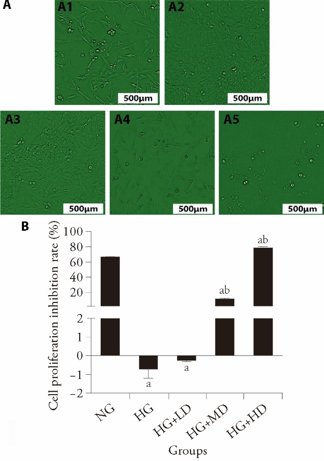
Figure 3 High-dose AC treatment showed significant inhibition of cell proliferation in vitro A: SV40-MES-13 cells were exposed to normal- or high-glucose conditions at 48 h at different concentrations of AC treatment (200 × with 15 × zoom); A1: NG; A2: HG; A3: HG + LD; A4: HG + MD; A5: HG + HD; B: inhibition rates of SV40-MES-13 cell proliferation in various groups were assayed by CCK8. NG: normal glucose, 5 mmol/L; HG: high glucose, 30 mmol/L; LD: low dose AC, 0.1 mmol/L; MD: medium dose AC, 1 mmol/L; HD: high dose AC, 10 mmol/L. AC: asiaticoside; LD: low-dose; MD: medium-dose; HD: high-dose: HG: high glucose. Differences among groups were assessed using one-way analysis of variance and the Bonferroni test. Data are presented as mean ± standard deviation (n = 5). aP < 0.01, compared with NG; bP < 0.01, compared with HG.
Figure 3 High-dose AC treatment showed significant inhibition of cell proliferation in vitro A: SV40-MES-13 cells were exposed to normal- or high-glucose conditions at 48 h at different concentrations of AC treatment (200 × with 15 × zoom); A1: NG; A2: HG; A3: HG + LD; A4: HG + MD; A5: HG + HD; B: inhibition rates of SV40-MES-13 cell proliferation in various groups were assayed by CCK8. NG: normal glucose, 5 mmol/L; HG: high glucose, 30 mmol/L; LD: low dose AC, 0.1 mmol/L; MD: medium dose AC, 1 mmol/L; HD: high dose AC, 10 mmol/L. AC: asiaticoside; LD: low-dose; MD: medium-dose; HD: high-dose: HG: high glucose. Differences among groups were assessed using one-way analysis of variance and the Bonferroni test. Data are presented as mean ± standard deviation (n = 5). aP < 0.01, compared with NG; bP < 0.01, compared with HG.
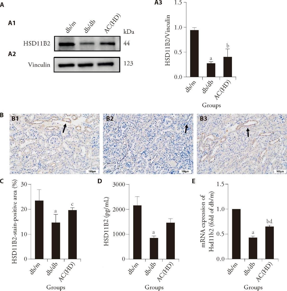
Figure 4 High-dose AC intervention protected against DKD mice by upregulating HSD11B2 A: the levels of HSD11B2 protein in mice with or without DKD as well as the intervention of high-dose AC revealed from Western blot; A1: representative western blot bands of HSD11B2 in each group; A2: representative western blot bands of Vinculin in each group; A3: quantitative analysis of HSD11B2 protein expression normalized to Vinculin; B: IHC-stained sites and relevant statistical analysis of HSD11B2 protein in kidney tissues; B1: db/m group; B2: db/db group; B3: AC (HD) group; C: HSD11B2-stain-positive area; D: ELISA results of HSD11B2 protein in the 3 groups; E: mRNA level of the expression of Hsd11b2 in the 3 groups. Mice were randomly divided into six groups and treated via oral gavage once daily for 12 weeks: control group (db/m), model group (db/db), low-dose AC group (5 g/kg), medium-dose AC group (15 g/kg), high-dose AC group (45 g/kg), and positive control group (valsartan, 5 mg/kg). All compounds were prepared as 0.2 mL suspensions for daily oral administration. AC: asiaticoside; DKD: diabetic kidney disease; ELISA: enzyme-linked immunosorbent assay; HSD11B2: 11-beta hydroxysteroid dehydrogenase type 2; IHC: immunohistochemistry. Black arrows indicate the distal convoluted tubules with positive staining. Differences among groups were assessed using one-way analysis of variance and the Bonferroni test. Data are presented as mean ± standard deviation (n = 5). aP < 0.01, compared with the db/m group; bP < 0.01, cP < 0.05,compared with the db/db group; dP < 0.01, AC (HD) compared with db/m group.
Figure 4 High-dose AC intervention protected against DKD mice by upregulating HSD11B2 A: the levels of HSD11B2 protein in mice with or without DKD as well as the intervention of high-dose AC revealed from Western blot; A1: representative western blot bands of HSD11B2 in each group; A2: representative western blot bands of Vinculin in each group; A3: quantitative analysis of HSD11B2 protein expression normalized to Vinculin; B: IHC-stained sites and relevant statistical analysis of HSD11B2 protein in kidney tissues; B1: db/m group; B2: db/db group; B3: AC (HD) group; C: HSD11B2-stain-positive area; D: ELISA results of HSD11B2 protein in the 3 groups; E: mRNA level of the expression of Hsd11b2 in the 3 groups. Mice were randomly divided into six groups and treated via oral gavage once daily for 12 weeks: control group (db/m), model group (db/db), low-dose AC group (5 g/kg), medium-dose AC group (15 g/kg), high-dose AC group (45 g/kg), and positive control group (valsartan, 5 mg/kg). All compounds were prepared as 0.2 mL suspensions for daily oral administration. AC: asiaticoside; DKD: diabetic kidney disease; ELISA: enzyme-linked immunosorbent assay; HSD11B2: 11-beta hydroxysteroid dehydrogenase type 2; IHC: immunohistochemistry. Black arrows indicate the distal convoluted tubules with positive staining. Differences among groups were assessed using one-way analysis of variance and the Bonferroni test. Data are presented as mean ± standard deviation (n = 5). aP < 0.01, compared with the db/m group; bP < 0.01, cP < 0.05,compared with the db/db group; dP < 0.01, AC (HD) compared with db/m group.
| 1. | Magliano DJ, Boyko EJ, committee IDFDAtes. IDF Diabetes Atlas. Brussels: International Diabetes Federation, 2021: 4-12. |
| 2. |
Cheng HT, Xu X, Lim PS, Hung KY. Worldwide epidemiology of diabetes-related end-stage renal disease, 2000-2015. Diabetes Care 2021; 44: 89-97.
DOI URL |
| 3. |
Cooper M, Warren AM. A promising outlook for diabetic kidney disease. Nat Rev Nephrol 2019; 15: 68-70.
DOI PMID |
| 4. | Yin B, Bi YM, Fan GJ, Xia YQ. Molecular mechanism of the effect of Huanglian Jiedu decoction on type 2 diabetes mellitus based on network pharmacology and molecular docking. J Diabetes Res 2020; 2020: 5273914. |
| 5. |
Shen S, Zhong H, Zhou X, et al. Advances in Traditional Chinese Medicine research in diabetic kidney disease treatment. Pharm Biol 2024; 62: 222-32.
DOI PMID |
| 6. |
Sari DCR, Budiharjo S, Afifah H, et al. Centella asiatica extract attenuates kidney fibrosis through reducing mesenchymal transition and inflammation in ureteral ligation model in mice. Front Pharmacol 2021; 12: 621894.
DOI URL |
| 7. |
Arfian N, Setyaningsih WAW, Anggorowati N, Romi MM, Sari DCR. Ethanol extract of Centella asiatica (Gotu Kola) attenuates tubular injury through inhibition of inflammatory cytokines and enhancement of anti-fibrotic factor in mice with 5/6 subtotal nephrectomy. Malays J Med Sci 2019; 26: 53-63.
DOI URL |
| 8. |
Masola B, Oguntibeju OO, Oyenihi AB. Centella asiatica ameliorates diabetes-induced stress in rat tissues via influences on antioxidants and inflammatory cytokines. Biomed Pharmacother 2018; 101: 447-57.
DOI URL |
| 9. |
Pittella F, Dutra RC, Junior DD, Lopes MTP, Barbosa NR. Antioxidant and cytotoxic activities of Centella asiatica (L) Urb. Int J Mol Sci 2009; 10: 3713-21.
DOI PMID |
| 10. |
Xu J, Wang S, Feng T, Chen Y, Yang G. Hypoglycemic and hypolipidemic effects of total saponins from Stauntonia chinensis in diabetic db/db mice. J Cell Mol Med 2018; 22: 6026-38.
DOI PMID |
| 11. |
Wang Z, Liu J, Sun W. Effects of asiaticoside on levels of podocyte cytoskeletal proteins and renal slit diaphragm proteins in adriamycin-induced rat nephropathy. Life Sci 2013; 93: 352-8.
DOI PMID |
| 12. |
Tang S, Xie X, Wang M, Yang L, Wei W. Protective effects of asiaticoside on renal ischemia reperfusion injury in vivo and in vitro. Bioengineered 2022; 13: 10235-43.
DOI URL |
| 13. |
Lu JZ, Ye D, Ma BL. Constituents, pharmacokinetics, and pharmacology of Gegen-Qinlian decoction. Front Pharmacol 2021; 12: 668418.
DOI URL |
| 14. | Ru J, Li P, Wang J, et al. TCMSP: A database of systems pharmacology for drug discovery from herbal medicines. J Cheminform 2014; 6: 13. |
| 15. |
Inderbinen SG, Zogg M, Kley M, Smieško M, Odermatt A. Species-specific differences in the inhibition of 11β-hydroxysteroid dehydrogenase 2 by itraconazole and posaconazole. Toxicol Appl Pharmacol 2021; 412: 115387.
DOI URL |
| 16. |
Jiang J, Yin J, Liu X, Wang H, Lu G. Erzhi formula extracts reverse renal injury in diabetic nephropathy rats by protecting the renal podocytes. Evid Based Complement Alternat Med 2018; 2018: 1741924.
DOI URL |
| 17. |
Zhu Q, Zeng J, Li J, et al. Effects of compound centella on oxidative stress and Keap1-Nrf2-ARE pathway expression in diabetic kidney disease rats. Evid Based Complement Alternat Med 2020; 2020: 9817932.
DOI URL |
| 18. | Sanaei-Ardekani M, Kamal S, Handy W, et al. Suppression of collagen Ⅳ alpha-2 subunit by prolyl hydroxylase domain inhibition via hypoxia-inducible factor-1 in chronic kidney disease. Pharmacol Res Perspect 2021; 9: e00872. |
| 19. |
Khawaja N, Abu-Shennar J, Saleh M, Dahbour SS, Khader YS, Ajlouni KM. The prevalence and risk factors of peripheral neuropathy among patients with type 2 diabetes mellitus; the case of Jordan. Diabetol Metab Syndr 2018; 10: 8.
DOI PMID |
| 20. |
Welsh GI, Hale LJ, Eremina V, et al. Insulin signaling to the glomerular podocyte is critical for normal kidney function. Cell Metab 2010; 12: 329-40.
DOI PMID |
| 21. |
Ghosh P, Sahoo R, Vaidya A, Chorev M, Halperin JA. Role of complement and complement regulatory proteins in the complications of diabetes. Endocr Rev 2015; 36: 272-88.
DOI PMID |
| 22. |
Zhu Q, Li XH, Chen HY, Jin QY. The effects of compound centella formula on oxinflammation and silent information regulator 1 in a high-fat diet/streptozotocin-induced diabetic kidney disease rat model. Exp Ther Med 2021; 22: 962.
DOI PMID |
| 23. |
Maulidiani, Abas F, Khatib A, et al. Metabolic alteration in obese diabetes rats upon treatment with Centella asiatica extract. J Ethnopharmacol 2016; 180: 60-9.
DOI PMID |
| 24. | Zhang Z, Ma J, Feng R, Wang Z. Centella asiatica inhibits renal interstitial fibrosis by regulating Smad3 and Smad7 expression in the TGFβ signaling pathway. Int J Clin Exp Pathol 2018; 11: 1009-17. |
| 25. | Jin L, Zheng D, Yang G, et al. Tilapia skin peptides ameliorate diabetic nephropathy in STZ-induced diabetic rats and HG-induced GMCs by improving mitochondrial dysfunction. Mar Drugs 2020: 18. |
| 26. |
Alshahrani S. Renin-angiotensin-aldosterone pathway modulators in chronic kidney disease: a comparative review. Front Pharmacol 2023; 14: 1101068.
DOI URL |
| 27. |
Homma M, Tanaka A, Hino K, et al. Assessing systemic 11β-hydroxysteroid dehydrogenase with serum cortisone/cortisol ratios in healthy subjects and patients with diabetes mellitus and chronic renal failure. Metabolism 2001; 50: 801-4.
DOI URL |
| 28. |
Hunter RW, Ivy JR, Flatman PW, et al. Hypertrophy in the distal convoluted tubule of an 11β-hydroxysteroid dehydrogenase type 2 knockout model. J Am Soc Nephrol 2015; 26: 1537-48.
DOI PMID |
| 29. |
Evans LC, Livingstone DE, Kenyon CJ, et al. A urine-concentrating defect in 11β-hydroxysteroid dehydrogenase type 2 null mice. Am J Physiol Renal Physiol 2012; 303: F494-502.
DOI URL |
| 30. |
Mune T, Suwa T, Morita H, et al. Longer HSD11B2 CA-repeat in impaired glucose tolerance and type 2 diabetes. Endocr J 2013; 60: 671-8.
DOI URL |
| 31. | Lavery GG, McTernan CL, Bain SC, Chowdhury TA, Hewison M, Stewart PM. Association studies between the HSD11B2 gene (encoding human 11β-hydroxysteroid dehydrogenase type 2), type 1 diabetes mellitus and diabetic nephropathy. Eur J Endocrinol 2002; 146: 553-8. |
| Viewed | ||||||
|
Full text |
|
|||||
|
Abstract |
|
|||||