Journal of Traditional Chinese Medicine ›› 2022, Vol. 42 ›› Issue (3): 379-388.DOI: 10.19852/j.cnki.jtcm.2022.03.005
• Research Article • Previous Articles Next Articles
Electroacupuncture preconditioning alleviates myocardial ischemia-reperfusion injury through the hypothalamic paraventricular nucleus- interposed nucleus nerve pathway
WEI Xiaotong1, LI Liaoyuan1, ZHANG Yating1, SHU Qi1, WANG Shuaiya1, CHEN Pianpian1, HU Ling2,3, YU Qing2,3( ), CAI Ronglin2,3()
- 1 Graduate School of Anhui University of Chinese Medicine, Hefei 230012, China
2 Institute of Acupuncture and Meridian, Anhui University of Chinese Medicine, Hefei 230012, China
3 Key Laboratory of Acupuncture and Moxibustion Fundamentals and Techniques of Anhui Province, Anhui University of Chinese Medicine, Hefei 230038, China
-
Received:2021-12-12Accepted:2022-02-22Online:2022-06-15Published:2022-05-20 -
Contact:YU Qing,CAI Ronglin -
About author:Prof. CAI Ronglin, Institute of Acupuncture and Meridian, Anhui University of Chinese Medicine, Hefei 230012, China. ronglincai@163.com;
-
Supported by:National Natural Science Foundation of China: Mechanism of GABA/Glu Neural Circuit in Lateral Hypothalamus-Parietal Nucleus in Alleviating Myocardial Ischemia-Reperfusion Injury by Acupuncture Preconditioning(82074536);Study on the Protective Effect of Acupuncture Pretreatment on Myocardial Ischemia-Reperfusion Injury Based on Hypothalamic-Cerebellar Neural Circuit(81774414);Mechanism of GABA Neural Circuit in the Paraventricular Nucleus of Hypothalamus and Ventrolateral Region of Medulla Oblongata in Alleviating Myocardial Ischemia-Reperfusion Injury Induced by Acupuncture Pretreatment(82104999);Natural Science Foundation of Anhui Province the Central Regulatory Mechanism of Acupuncture Regulating Cardiac Function(2108085Y30);Anhui Province University Outstanding Top Talent Cultivation Funding Project(gxgwfx2019025)
Cite this article
WEI Xiaotong, LI Liaoyuan, ZHANG Yating, SHU Qi, WANG Shuaiya, CHEN Pianpian, HU Ling, YU Qing, CAI Ronglin. Electroacupuncture preconditioning alleviates myocardial ischemia-reperfusion injury through the hypothalamic paraventricular nucleus- interposed nucleus nerve pathway[J]. Journal of Traditional Chinese Medicine, 2022, 42(3): 379-388.
share this article

Figure 1 Changes of ST segment in each group at different time periods A: sham group before ligation; B: sham group received an empty needle; C: sham group was vacuumed for 2 h; D: MIRI group before ligation; E: MIRI group was ligation for 30 min; F: MIRI group was reperfused for 2 h; G: EA group before ligation; H: EA group was ligation for 30 min; I: EA group was reperfused for 2 h; J: lesion +EA group before ligation; K: lesion +EA group was ligation for 30 min L. Lesion + EA group was reperfused for 2 h. The MIRI group refers to the myocardial ischemia reperfusion injury model group. EA group refers to the electroacupuncture of the heart meridians group + myocardial ischemia-reperfusion injury group. The lesion + EA group refers to lesion of the intercerebellar nucleus + electroacupuncture pretreatment + MIRI group. PVN: paraventricular nucleus; MIRI: myocardial ischemia-reperfusion injury; EA: electroacupuncture.
Figure 1 Changes of ST segment in each group at different time periods A: sham group before ligation; B: sham group received an empty needle; C: sham group was vacuumed for 2 h; D: MIRI group before ligation; E: MIRI group was ligation for 30 min; F: MIRI group was reperfused for 2 h; G: EA group before ligation; H: EA group was ligation for 30 min; I: EA group was reperfused for 2 h; J: lesion +EA group before ligation; K: lesion +EA group was ligation for 30 min L. Lesion + EA group was reperfused for 2 h. The MIRI group refers to the myocardial ischemia reperfusion injury model group. EA group refers to the electroacupuncture of the heart meridians group + myocardial ischemia-reperfusion injury group. The lesion + EA group refers to lesion of the intercerebellar nucleus + electroacupuncture pretreatment + MIRI group. PVN: paraventricular nucleus; MIRI: myocardial ischemia-reperfusion injury; EA: electroacupuncture.
Table 1 Statistical analysis of electrocardiogram of rats in each group (`x ± s)
| Group | n | Before ligation | Ligation for 30 min | Reperfusion 2 h |
|---|---|---|---|---|
| Sham | 6 | 0.07±0.05 | 0.10±0.10 | 0.07±0.02 |
| MIRI | 6 | 0.10±0.04 | 0.62±0.17ad | 0.28±0.07bd |
| EA | 6 | 0.07±0.03 | 0.31±0.05aef | 0.14±0.04cef |
| Lesion + EA | 6 | 0.10±0.05 | 0.45±0.13a | 0.21±0.04c |
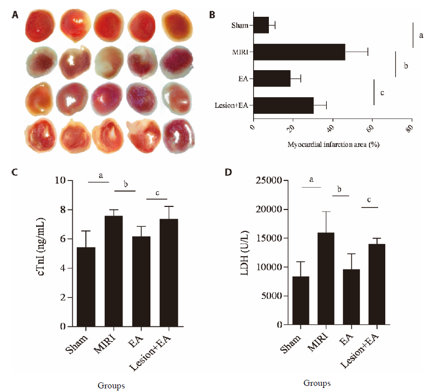
Figure 2 Statistical analysis of myocardial infarction size, cTnI and LDH between groups A: myocardial infarction area of each group (red is normal myocardial tissue, white is infarct area); B: percentage of myocardial infarction area in each group; C, D: comparison of cTnI and LDH in each group. The MIRI group refers to the myocardial ischemia reperfusion injury model group, EA group refers to the electroacupuncture of the heart meridians group + myocardial ischemia-reperfusion injury group, the lesion + EA group refers to lesion of the intercerebellar nucleus + electroacupuncture pretreatment + MIRI group. PVN: paraventricular nucleus; MIRI: myocardial ischemia-reperfusion injury; EA: electroacupuncture; cTnI: cardiac troponin I; LDH: Lactate dehydrogenase. Compared with the sham group, aP < 0.01; compared with the MIRI group, bP < 0.01 and compared with the lesion + EA group, cP < 0.05.
Figure 2 Statistical analysis of myocardial infarction size, cTnI and LDH between groups A: myocardial infarction area of each group (red is normal myocardial tissue, white is infarct area); B: percentage of myocardial infarction area in each group; C, D: comparison of cTnI and LDH in each group. The MIRI group refers to the myocardial ischemia reperfusion injury model group, EA group refers to the electroacupuncture of the heart meridians group + myocardial ischemia-reperfusion injury group, the lesion + EA group refers to lesion of the intercerebellar nucleus + electroacupuncture pretreatment + MIRI group. PVN: paraventricular nucleus; MIRI: myocardial ischemia-reperfusion injury; EA: electroacupuncture; cTnI: cardiac troponin I; LDH: Lactate dehydrogenase. Compared with the sham group, aP < 0.01; compared with the MIRI group, bP < 0.01 and compared with the lesion + EA group, cP < 0.05.
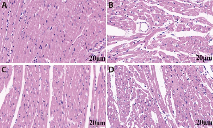
Figure 3 Hematoxylin-eosin staining results in each group A: sham group; B: MIRI group; C: EA group; D: lesion + EA group. The MIRI group refers to the myocardial ischemia reperfusion injury model group, EA group refers to the electroacupuncture of the heart meridians group + myocardial ischemia-reperfusion injury group, the lesion + EA group refers to lesion of the intercerebellar nucleus + electroacupuncture pretreatment + MIRI group. PVN: paraventricular nucleus; MIRI: myocardial ischemia-reperfusion injury; EA: electroacupuncture. Partial image is 400 times, scale is 20 μm.
Figure 3 Hematoxylin-eosin staining results in each group A: sham group; B: MIRI group; C: EA group; D: lesion + EA group. The MIRI group refers to the myocardial ischemia reperfusion injury model group, EA group refers to the electroacupuncture of the heart meridians group + myocardial ischemia-reperfusion injury group, the lesion + EA group refers to lesion of the intercerebellar nucleus + electroacupuncture pretreatment + MIRI group. PVN: paraventricular nucleus; MIRI: myocardial ischemia-reperfusion injury; EA: electroacupuncture. Partial image is 400 times, scale is 20 μm.
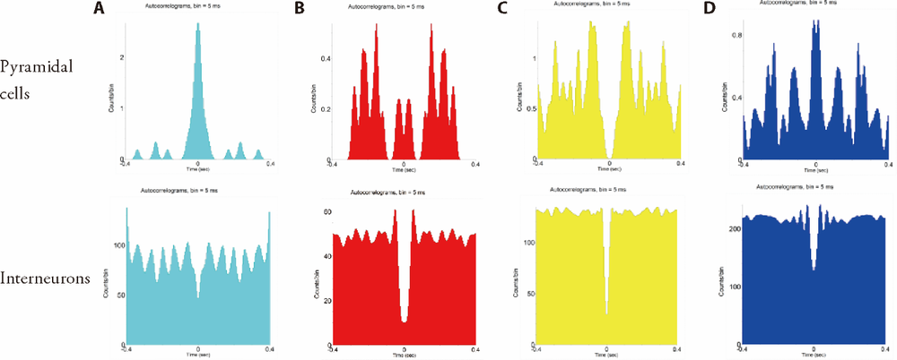
Figure 4 Autocorrelation analysis of PVN neuron electrical activity in each group of rats A: sham group; B: MIRI group; C: EA group; D: lesion + EA group. The MIRI group refers to the myocardial ischemia reperfusion injury model group, EA group refers to the electroacupuncture of the heart meridians group + myocardial ischemia-reperfusion injury group, the lesion +EA group refers to lesion of the intercerebellar nucleus + electroacupuncture pretreatment + MIRI group. PVN: paraventricular nucleus; MIRI: myocardial ischemia-reperfusion injury; EA: electroacupuncture.
Figure 4 Autocorrelation analysis of PVN neuron electrical activity in each group of rats A: sham group; B: MIRI group; C: EA group; D: lesion + EA group. The MIRI group refers to the myocardial ischemia reperfusion injury model group, EA group refers to the electroacupuncture of the heart meridians group + myocardial ischemia-reperfusion injury group, the lesion +EA group refers to lesion of the intercerebellar nucleus + electroacupuncture pretreatment + MIRI group. PVN: paraventricular nucleus; MIRI: myocardial ischemia-reperfusion injury; EA: electroacupuncture.
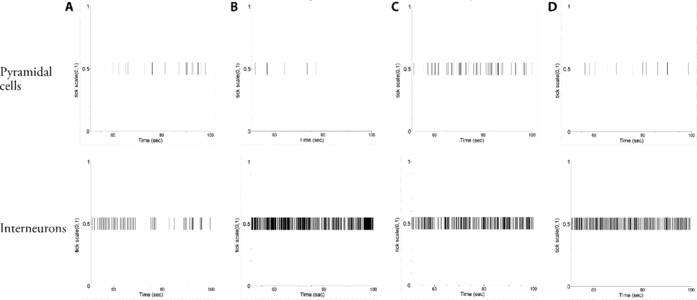
Figure 5 Comparison of raster images of PVN neuron electrical activity in rats from each group A: sham group; B: MIRI group; C: EA group; D: lesion + EA group. The MIRI group refers to the myocardial ischemia reperfusion injury model group, EA group refers to the electroacupuncture of the heart meridians group + myocardial ischemia-reperfusion injury group, the lesion +EA group refers to lesion of the intercerebellar nucleus + electroacupuncture pretreatment + MIRI group. PVN: paraventricular nucleus; MIRI: myocardial ischemia-reperfusion injury; EA: electroacupuncture.
Figure 5 Comparison of raster images of PVN neuron electrical activity in rats from each group A: sham group; B: MIRI group; C: EA group; D: lesion + EA group. The MIRI group refers to the myocardial ischemia reperfusion injury model group, EA group refers to the electroacupuncture of the heart meridians group + myocardial ischemia-reperfusion injury group, the lesion +EA group refers to lesion of the intercerebellar nucleus + electroacupuncture pretreatment + MIRI group. PVN: paraventricular nucleus; MIRI: myocardial ischemia-reperfusion injury; EA: electroacupuncture.
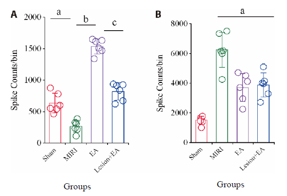
Figure 6 PVN neuron spike counts in each group. A: number of firings of pyramidal cells; B: number of firings of interneurons. The MIRI group refers to the myocardial ischemia reperfusion injury model group, EA group refers to the electroacupuncture of the heart meridians group + myocardial ischemia-reperfusion injury group, the lesion + EA group refers to lesion of the intercerebellar nucleus + electroacupuncture pretreatment + MIRI group. PVN: paraventricular nucleus; MIRI: myocardial ischemia-reperfusion injury; EA: electroacupuncture. Compared with the sham group, aP < 0.01; compared with the MIRI group, bP < 0.01; compared with the lesion+ EA group, cP < 0.01.
Figure 6 PVN neuron spike counts in each group. A: number of firings of pyramidal cells; B: number of firings of interneurons. The MIRI group refers to the myocardial ischemia reperfusion injury model group, EA group refers to the electroacupuncture of the heart meridians group + myocardial ischemia-reperfusion injury group, the lesion + EA group refers to lesion of the intercerebellar nucleus + electroacupuncture pretreatment + MIRI group. PVN: paraventricular nucleus; MIRI: myocardial ischemia-reperfusion injury; EA: electroacupuncture. Compared with the sham group, aP < 0.01; compared with the MIRI group, bP < 0.01; compared with the lesion+ EA group, cP < 0.01.

Figure 7 Comparison of spectral energy diagrams between the groups (time = 300 s; frequency = 40 Hz). A: sham group; B: MIRI group; C: EA group; D: lesion + EA group. The MIRI group refers to the myocardial ischemia reperfusion injury model group, EA group refers to the electroacupuncture of the heart meridians group + myocardial ischemia-reperfusion injury group, the lesion + EA group refers to lesion of the intercerebellar nucleus + electroacupuncture pretreatment + MIRI group. PVN: paraventricular nucleus; MIRI: myocardial ischemia-reperfusion injury; EA: electroacupuncture.
Figure 7 Comparison of spectral energy diagrams between the groups (time = 300 s; frequency = 40 Hz). A: sham group; B: MIRI group; C: EA group; D: lesion + EA group. The MIRI group refers to the myocardial ischemia reperfusion injury model group, EA group refers to the electroacupuncture of the heart meridians group + myocardial ischemia-reperfusion injury group, the lesion + EA group refers to lesion of the intercerebellar nucleus + electroacupuncture pretreatment + MIRI group. PVN: paraventricular nucleus; MIRI: myocardial ischemia-reperfusion injury; EA: electroacupuncture.

Figure 8 c-fos expression in the PVN of each group (immunofluorescence staining, partial image was 200 times, scale was 50 μm) A: sham group; B: MIRI group; C: EA group; D: lesion + EA group. The MIRI group refers to the myocardial ischemia reperfusion injury model group, EA group refers to the electroacupuncture of the heart meridians group + myocardial ischemia-reperfusion injury group, the lesion + EA group refers to lesion of the intercerebellar nucleus + electroacupuncture pretreatment + MIRI group. PVN: paraventricular nucleus; MIRI: myocardial ischemia-reperfusion injury; EA: electroacupuncture.
Figure 8 c-fos expression in the PVN of each group (immunofluorescence staining, partial image was 200 times, scale was 50 μm) A: sham group; B: MIRI group; C: EA group; D: lesion + EA group. The MIRI group refers to the myocardial ischemia reperfusion injury model group, EA group refers to the electroacupuncture of the heart meridians group + myocardial ischemia-reperfusion injury group, the lesion + EA group refers to lesion of the intercerebellar nucleus + electroacupuncture pretreatment + MIRI group. PVN: paraventricular nucleus; MIRI: myocardial ischemia-reperfusion injury; EA: electroacupuncture.
| 1 | Hao F, Cai RL, Yu Q, et al. Effect of electroacupuncture preconditioning on the expressions of NF-κB p65, IκBα and IKKβ in myocardial tissue of the rats with acute myocardial ischemia-reperfusion injury. Zhong Guo Zhen Jiu 2020; 40:1103-7. |
| 2 |
Yang L, Yang J, Wang Q, et al. Cardioprotective effects of electroacupuncture pretreatment on patients undergoing heart valve replacement surgery: a randomized controlled trial. Ann Thorac Surg 2010; 89:781-6.
DOI URL |
| 3 |
Jang I, Cho K, Moon S, et al. A study on the central neural pathway of the heart, Nei-Kuan (EH-6) and Shen-Men (He-7) with neural tracer in rats. Am J Chin Med 2003; 31:591-609.
DOI URL |
| 4 | Chi HJ, Chen ML, Yang XC, et al. Progress in therapies for myocardial ischemia reperfusion injury. Curr Drug Targets 2017; 18:1712-21. |
| 5 |
Zhong MK, Duan YC, Chen AD, et al. Paraventricular nucleus is involved in the central pathway of cardiac sympathetic afferent reflex in rats. Exp Physiol 2008; 93:746-53.
DOI URL |
| 6 |
Chen WW, Xiong XQ, Chen Q, et al. Cardiac sympathetic afferent reflex and its implications for sympathetic activation in chronic heart failure and hypertension. Acta Physiol (Oxf) 2015; 213:778-94.
DOI PMID |
| 7 | Chen C, Zhang Y, Cheng XY, et al. Central nervous system regulation mechanism of myocardial ischemia-reperfusion injury in rats: neuronal excitability in the paraventricular nucleus of the hypothalamus. Zhong Hua Ma Zui Xue Za Zhi 2018; 38:1293-7. |
| 8 | Cai RL, Cui S, Wu ZJ, et al. Effect of electroacupuncture at “Shenmen” (HT7)-“Tongli” (HT5) of heart meridian on neuronal activities in paraventricular nucleus of hypothalamus in myo-cardial ischemia rats. Zhen Ci Yan Jiu 2018; 43:406-13. |
| 9 |
Zhu JN, Yung WH, Kwok-Chong Chow B, et al. The cerebellar-hypothalamic circuits: potential pathways underlying cerebellar involvement in somatic-visceral integration. Brain Res Rev 2006; 52:93-106.
DOI URL |
| 10 |
Lu JH, Mao HN, Cao BB, et al. Effect of cerebellohypothalamic glutamatergic projections on immune function. Cerebellum 2012; 11:905-16.
DOI URL |
| 11 | Han J, Xuan JL, Hu HR, et al. Protective effect against myocardial ischemia reperfusion injuries induced by hyperoside preconditioning and its relationship with PI3K/Akt signaling pathway in rats. Zhong Guo Zhong Yao Za Zhi 2015; 40:118-23. |
| 12 | Paxinos G, Watson C. The rat brain in stereotaxic coordinates. People's Medical Publishing House, 2005, in press. |
| 13 | Liu F, Li BM. Brain function damage and inactivation methods commonly used in learning and memory research. Zhong Guo Xing Wei Yi Xue Ke Xue 2006; 222-3. |
| 14 | Lin WZ, Wang P. Experimental acupuncture and moxibustion. Shanghai Science and Technology Press, 1999, in press. |
| 15 |
Barthó P, Hirase H, Monconduit L, et al. Characterization of neo-cortical principal cells and interneurons by network inter-actions and extracellular features. J Neurophysiol 2004; 92:600-8.
PMID |
| 16 | Xu JM, Wang CQ, Lin LN. Multi-channel in vivo recording techniques: signal processing of action potentials and local field potentials. Sheng Li Xue Bao 2014; 66:349-57. |
| 17 | Painovich J, Longhurst J. Integrating acupuncture into the cardio-logy clinic: can it play a role? Sheng Li Xue Bao 2015; 67:19-31. |
| 18 |
Wang JS, Yu XD, Deng S, et al. Acupuncture on treating angina pectoris: a systematic review. Medicine 2020; 99:e18548.
DOI URL |
| 19 | Cui S, Xu J, Wang J, et al. Effect of electroacupuncture stimulation of heart meridian on autonomic nervous activities in acute myocardial ischemia rats. Zhen Ci Yan Jiu 2016; 41:515-20. |
| 20 |
Armstrong K, Gokal R, Todorsky W. Neuromodulating influence of two electroacupuncture treatments on heart rate variability, stress, and vagal activity. J Altern Complement Med 2020; 26:928-36.
DOI URL |
| 21 | Sun YZ, Yao J, Zhou L. Research progress in the ancient and modern methods of acupuncture method of matching points of the original collaterals. Liaoning Zhong Yi Yao Da Xue Xue Bao 2019; 21:5-9. |
| 22 |
Cui S, Wang K, Wu SB, et al. Electroacupuncture modulates the activity of the hippocampus-nucleus tractus solitarius-vagus nerve pathway to reduce myocardial ischemic injury. Neural Regen Res 2018; 13:1609-18.
DOI URL |
| 23 | Zhang HH, Wang YJ, Zheng C, et al. Apelin in the hypothalamic paraventricular nucleus improves cardiac function in surgical trauma rats. Sheng Li Xue Bao 2018; 70:99-105. |
| 24 |
Ciriello J, Kline RL, Zhang TX, et al. Lesions of the paraventricular nucleus alter the development of spontaneous hypertension in the rat. Brain Res 1984; 310:355-9.
PMID |
| 25 |
Xu B, Zheng H, Patel KP. Relative contributions of the thalamus and the paraventricular nucleus of the hypothalamus to the cardiac sympathetic afferent reflex. Am J Physiol Regul Integr Comp Physiol 2013; 305:R50-9.
DOI URL |
| 26 |
Saab CY, Willis WD. Cerebellar stimulation modulates the intensity of a visceral nociceptive reflex in the rat. Exp Brain Res 2002; 146:117-21.
DOI PMID |
| 27 |
Onat F, Cavdar S. Cerebellar connections: hypothalamus. Cerebellum 2003; 2:263-9.
DOI URL |
| 28 | Kullmann S, Veit R. Resting-state functional connectivity of the human hypothalamus. Handb Clin Neurol 2021; 179:113-24. |
| 29 |
Dietrichs E, Haines DE. Interconnections between hypothalamus and cerebellum. Anat Embryol 1989; 179:207-20.
PMID |
| 30 | Yu Q, Cai RL, Shao XF, et al. Effect of electroacupuncture preconditioning on the contents of dopamine and 5-hydroxytryptamine in lateral hypothalamus area and cerebellar fastigial nucleus of rats with myocardial ischemia-reperfusion injury. Zhong Guo Zhen Jiu 2021; 41:525-30. |
| 31 |
Wen YQ, Zhu JN, Zhang YP, et al. Cerebellar interpositus nuclear inputs impinge on paraventricular neurons of the hypothalamus in rats. Neurosci Lett 2004; 370:25-9.
DOI URL |
| 32 |
van den Hoogen NJ, Kwok CHT, Trang T. Identifying the neurodevelopmental differences of opioid withdrawal. Cell Mol Neurobiol 2021; 41:1145-55.
DOI PMID |
| 33 |
Coyle JT, Molliver ME, Kuhar MJ. In situ injection of kainic acid: a new method for selectively lesioning neural cell bodies while sparing axons of passage. J Comp Neurol 1978; 180:301-23.
PMID |
| Viewed | ||||||
|
Full text |
|
|||||
|
Abstract |
|
|||||