
Journal of Traditional Chinese Medicine ›› 2026, Vol. 46 ›› Issue (5): 1180-1193.DOI: 10.19852/j.cnki.jtcm.2026.05.012
• Original Articles • Previous Articles Next Articles
Jianpi Huayu decoction (健脾化瘀方) induces hepatocellular carcinoma apoptosis through flavonoid components targeting tumor protein p53 activation and serine-threonine protein kinase 1/B-cell lymphoma 2 inhibition: a network pharmacology-based study with experimental validation
CHEN Xiaokai1, XIE Yingjie2, WU Hui3, LI Yong4, LU Yantong5, TAN Wei6, WANG Changjun6( ), HUANG Xuhui6,7()
- 1
Department of Pharmacy ,Guangdong Provincial People's Hospital (Guangdong Academy of Medical Sciences), Southern Medical University Guangzhou 510000, China
2One Departments of Cadre Ward ,General Hospital of Southern Theater Command of Chinese PLA Guangzhou 510000, China
3South China University of Technology School of Medicine Guangzhou 510000, China
4Guangdong Provincial Key Laboratory for Research and Evaluation of Pharmaceutical Preparations ,Guangdong Pharmaceutical University Guangzhou 510000, China
5Science and Technology Innovation Center of Guangzhou University of Chinese Medicine ,Guangzhou University of Chinese Medicine Guangzhou 510000, China
6Department of Traditional Chinese Medicine ,Guangdong Geriatric institute, Guangdong Provincial People’s Hospital (Guangdong Academy of Medical Sciences), Southern Medical University Guangzhou 510000, China
7School of Traditional Chinese Medicine ,Southern Medical University Guangzhou 510000, China
-
Received:2025-05-30Accepted:2026-01-28Online:2026-10-15Published:2026-10-09 -
Contact:WANG Changjun, Department of Traditional Chinese Medicine, Guangdong Geriatric institute, Guangdong Provincial People’s Hospital (Guangdong Academy of Medical Sciences), Southern Medical University, Guangzhou 510000, China, gzwchj@126.com; HUANG Xuhui, Department of Traditional Chinese Medicine, Guangdong Geriatric institute, Guangdong Provincial People’s Hospital (Guangdong Academy of Medical Sciences), Southern Medical University, Guangzhou 510000, China; School of Traditional Chinese Medicine, Southern Medical University, Guangzhou 510000, China, api007@163.com,Telephone: +86-13544437648 -
About author:CHEN Xiaokai and XIE Yingjie are co-first authors and contributed equally to this work
-
Supported by:Natural Science Foundation of Guangdong Province: the Role of Exosomal MicroRNA-122/Transforming Growth Factor-β/Smad Pathway in Inducing Myeloid-Derived Suppressor Cell Differentiation and Promoting Liver Cancer Dormancy and the Intervention Study of Jianpi Huayu Decoction(2022A1515011260);Administration of Traditional Chinese Medicine of Guangdong Province: Investigation of the Inhibitory Effect and Intervention Mechanism of Jianpi Huayu decoction on Postoperative Recurrence of Liver Cancer via the Phosphoinositide 3-Kinase/ Serine-Threonine Protein Kinase 1/Nuclear Factor Kappa-B Signaling Pathway(20251002)

Cite this article
CHEN Xiaokai, XIE Yingjie, WU Hui, LI Yong, LU Yantong, TAN Wei, WANG Changjun, HUANG Xuhui. Jianpi Huayu decoction (健脾化瘀方) induces hepatocellular carcinoma apoptosis through flavonoid components targeting tumor protein p53 activation and serine-threonine protein kinase 1/B-cell lymphoma 2 inhibition: a network pharmacology-based study with experimental validation[J]. Journal of Traditional Chinese Medicine, 2026, 46(5): 1180-1193.
share this article
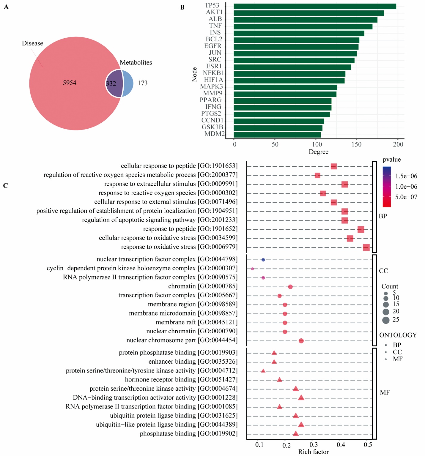
Figure 1 Network pharmacological analysis of the active components of JHD-containing serum A: venn diagram showing the intersection between potential target genes of JHD active components and HCC-related geness; B: bar chart of the top 20 intersecting targets ranked by degree value; C: GO enrichment analysis (Biological Process, Cellular Component, Molecular Function) of the top 50 intersecting targets, presented as bubble plots; PI3K: phosphatidylinositol 3-kinase; TP53: tumor protein 53; AKT1: serine-threonine kinase 1; ALB: albumin; TNF: tumor necrosis factor; INS: insulin; BCL2: B-cell lymphoma 2; EGFR: epidermal growth factor receptor; JUN: Jun proto-oncogene; SRC: SRC proto-oncogene; ESR1: estrogen receptor 1; NFKB1: nuclear factor kappa B subunit 1; HIF1A: hypoxia inducible factor 1 subunit alpha; MAPK3: mitogen-activated protein kinases 3; MMP9: matrix metallopeptidase 9; PPARG: peroxisome proliferator activated receptor gamma; IFNG: interferon gamma; PTGS2: prostaglandin-endoperoxide synthase 2; CCND1: cyclin d1; GSK3B: glycogen synthase kinase 3 beta; MDM2: MDM2 proto-oncogene; JHD: Jianpi Huayu decoction; GO: gene ontology; KEGG: kyoto encyclopedia of genes and genomes; BP: biological process; CC: cellular component; MF: molecular function.
Figure 1 Network pharmacological analysis of the active components of JHD-containing serum A: venn diagram showing the intersection between potential target genes of JHD active components and HCC-related geness; B: bar chart of the top 20 intersecting targets ranked by degree value; C: GO enrichment analysis (Biological Process, Cellular Component, Molecular Function) of the top 50 intersecting targets, presented as bubble plots; PI3K: phosphatidylinositol 3-kinase; TP53: tumor protein 53; AKT1: serine-threonine kinase 1; ALB: albumin; TNF: tumor necrosis factor; INS: insulin; BCL2: B-cell lymphoma 2; EGFR: epidermal growth factor receptor; JUN: Jun proto-oncogene; SRC: SRC proto-oncogene; ESR1: estrogen receptor 1; NFKB1: nuclear factor kappa B subunit 1; HIF1A: hypoxia inducible factor 1 subunit alpha; MAPK3: mitogen-activated protein kinases 3; MMP9: matrix metallopeptidase 9; PPARG: peroxisome proliferator activated receptor gamma; IFNG: interferon gamma; PTGS2: prostaglandin-endoperoxide synthase 2; CCND1: cyclin d1; GSK3B: glycogen synthase kinase 3 beta; MDM2: MDM2 proto-oncogene; JHD: Jianpi Huayu decoction; GO: gene ontology; KEGG: kyoto encyclopedia of genes and genomes; BP: biological process; CC: cellular component; MF: molecular function.
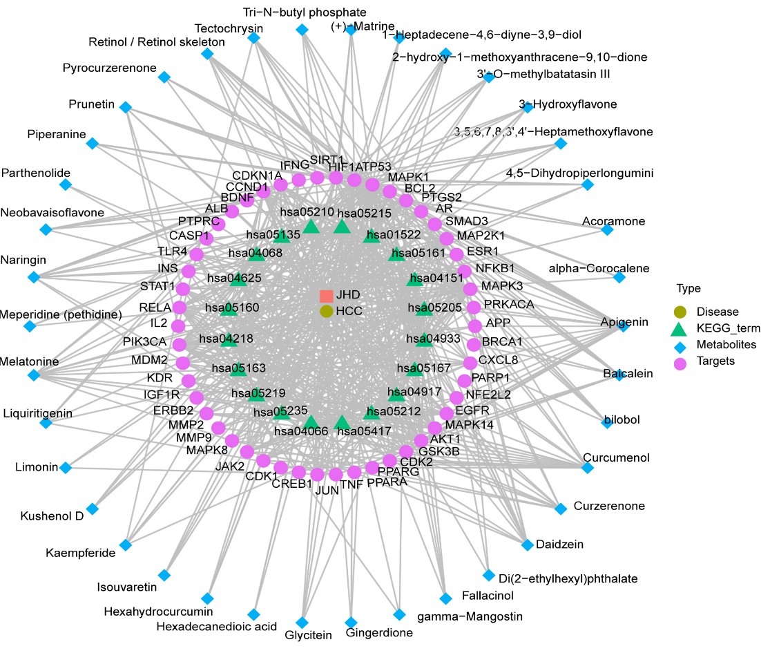
Figure 2 PPI and "component-target" interaction network analysis The “JHD-Blood Component-Target-Pathway-HCC” network, depicting the top 50 protein targets. Interaction analysis revealed that key serum components of JHD, apigenin, curcumenol, daidzein, naringin, and curzerenone, can act on critical targets including TP53, AKT1, ALB, TNF, INS, BCL2, EGFR, and JUN. In this network, square nodes represent chemical components, circular nodes represent protein targets, and connecting lines denote interactions between them. The number of lines originating from a component signifies its degree value, reflecting its broader influence on multiple targets. SIRT1: sirtuin 1; HIF1A: hypoxia inducible factor 1 subunit alpha; TP53: tumor protein P53; MAPK1: mitogen-activated protein kinase 1; PTGS2: prostaglandin-endoperoxide synthase 2; AR: androgen receptor; SMAD3: SMAD family member 3; MAP2K1: mitogen-activated protein kinase1; ESR1: estrogen receptor 1; NFKB1: nuclear factor kappa B subunit 1; MAPK3: mitogen-activated protein kinase 3; PRKACA: protein kinase cAMP-activated catalytic subunit alpha; APP: amyloid beta precursor protein; CXCL8: C-X-C motif chemokine ligand 8; PARP1: poly (ADP-ribose) polymerase 1; NFE2L2: NFE2 like bZIP transcription factor 2; EGFR: epidermal growth factor receptor; MAPK14: mitogen-activated protein kinase 14; AKT1: serine/threonine kinase 1; GSK3B: glycogen synthase kinase 3 beta; CDK2: cyclin dependent kinase 2; PPARG: peroxisome proliferator activated receptor gamma; PPARA: peroxisome proliferator activated receptor alpha; TNF: tumor necrosis factor; JUN: Jun proto-oncogene, AP-1 transcription factor subunit; CREB1: CAMP responsive element binding protein 1; CDK1: cyclin dependent kinase 1; JAK2: Janus kinase 2; MAPK8: mitogen-activated protein kinase 8; MMP9: matrix metallopeptidase 9; MMP2: matrix metallopeptidase 2; ERBB2: Erb-b2 receptor tyrosine kinase 2; IGF1R: insulin like growth factor 1 receptor; KDR: kinase insert domain receptor; PIK3CA: phosphatidylinositol-4,5-bisphosphate 3-kinase catalytic subunit alpha; IL-2: interleukin 2; RELA: RELA proto-oncogene, NF-kB subunit; STAT1: signal transducer and activator of transcription 1; INS: insulin; TLR4: toll like receptor 4; CASP1: caspase 1; PTPRC: protein tyrosine phosphatase receptor type C; ALB: Albumin; BDNF: brain derived neurotrophic factor; CCND1: cyclin D1; CDKN1A: cyclin dependent kinase inhibitor 1A; IFNG: interferon gamma; JHD: Jianpi Huayu decoction; HCC: hepatocellular carcinoma.
Figure 2 PPI and "component-target" interaction network analysis The “JHD-Blood Component-Target-Pathway-HCC” network, depicting the top 50 protein targets. Interaction analysis revealed that key serum components of JHD, apigenin, curcumenol, daidzein, naringin, and curzerenone, can act on critical targets including TP53, AKT1, ALB, TNF, INS, BCL2, EGFR, and JUN. In this network, square nodes represent chemical components, circular nodes represent protein targets, and connecting lines denote interactions between them. The number of lines originating from a component signifies its degree value, reflecting its broader influence on multiple targets. SIRT1: sirtuin 1; HIF1A: hypoxia inducible factor 1 subunit alpha; TP53: tumor protein P53; MAPK1: mitogen-activated protein kinase 1; PTGS2: prostaglandin-endoperoxide synthase 2; AR: androgen receptor; SMAD3: SMAD family member 3; MAP2K1: mitogen-activated protein kinase1; ESR1: estrogen receptor 1; NFKB1: nuclear factor kappa B subunit 1; MAPK3: mitogen-activated protein kinase 3; PRKACA: protein kinase cAMP-activated catalytic subunit alpha; APP: amyloid beta precursor protein; CXCL8: C-X-C motif chemokine ligand 8; PARP1: poly (ADP-ribose) polymerase 1; NFE2L2: NFE2 like bZIP transcription factor 2; EGFR: epidermal growth factor receptor; MAPK14: mitogen-activated protein kinase 14; AKT1: serine/threonine kinase 1; GSK3B: glycogen synthase kinase 3 beta; CDK2: cyclin dependent kinase 2; PPARG: peroxisome proliferator activated receptor gamma; PPARA: peroxisome proliferator activated receptor alpha; TNF: tumor necrosis factor; JUN: Jun proto-oncogene, AP-1 transcription factor subunit; CREB1: CAMP responsive element binding protein 1; CDK1: cyclin dependent kinase 1; JAK2: Janus kinase 2; MAPK8: mitogen-activated protein kinase 8; MMP9: matrix metallopeptidase 9; MMP2: matrix metallopeptidase 2; ERBB2: Erb-b2 receptor tyrosine kinase 2; IGF1R: insulin like growth factor 1 receptor; KDR: kinase insert domain receptor; PIK3CA: phosphatidylinositol-4,5-bisphosphate 3-kinase catalytic subunit alpha; IL-2: interleukin 2; RELA: RELA proto-oncogene, NF-kB subunit; STAT1: signal transducer and activator of transcription 1; INS: insulin; TLR4: toll like receptor 4; CASP1: caspase 1; PTPRC: protein tyrosine phosphatase receptor type C; ALB: Albumin; BDNF: brain derived neurotrophic factor; CCND1: cyclin D1; CDKN1A: cyclin dependent kinase inhibitor 1A; IFNG: interferon gamma; JHD: Jianpi Huayu decoction; HCC: hepatocellular carcinoma.
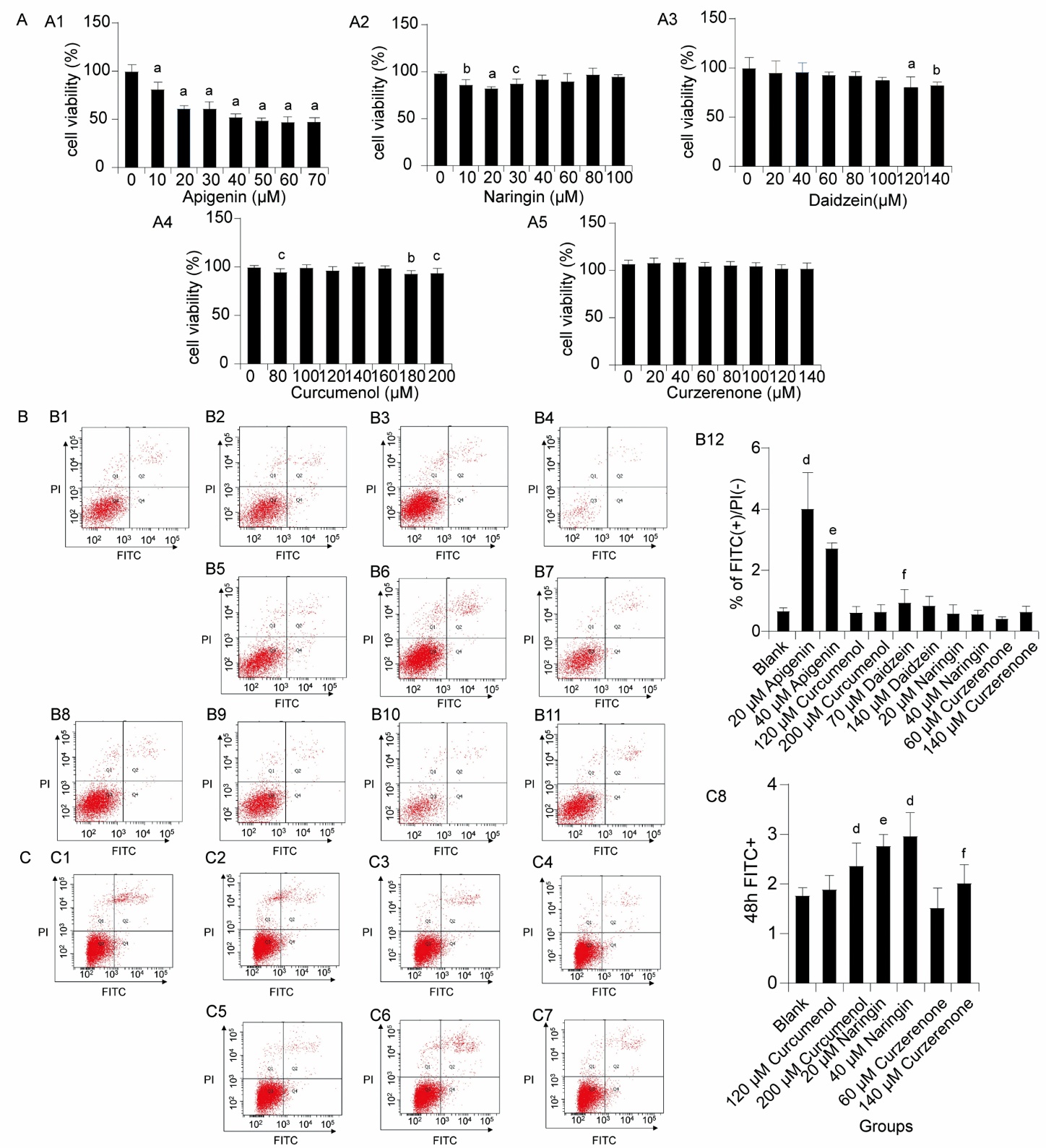
Figure 3 Apigenin, curcumenol, daidzein, naringin, and curzerenone promote apoptosis of HepG2 cells A: CCK-8 assay was employed to evaluate cell apoptosis; A1: bar chart of cell viability in the apigenin group; A2: bar chart of cell viability in the naringin group; A3: bar chart of cell viability in the daidzein group; A4: bar chart of cell viability in the curcumenol group; A5: bar chart of cell viability in the curzerenone group. B: Annexin V-FITC and PI staining was utilized to assess cell apoptosis via flow cytometry; B1: blank group; B2: 20 μM apigenin group; B3: 120 μM curcumenol group; B4: 70 μM daidzein group; B5: 40 μM apigenin group; B6: 200 μM curcumenol group; B7: 140 μM daidzein group; B8: 60 μM curzerenone group; B9: 140 μM curzerenone group; B10: 20 μM naringin group; B11: 40 μM naringin group; B12: bar gaph of statistics. C: Annexin V-FITC and PI staining was utilized to assess cell apoptosis via flow cytometry; C1: blank group; C2: 120 μM curcumenol group; C3: 20 μM naringin group; C4: 60 μM curzerenone group; C5: 200 μM curcumenol group; C6: 40 μM naringin group; C7: 140 μM curzerenone group; C8: bar gaph of statistics. For A: HepG2 cells were treated with apigenin, curcumenol, daidzein, naringin, and curzerenone for 24 h in vitro (n = 6); For B: HepG2 cells were exposed to these monomers for 24 h in vitro (n = 4); For C: HepG2 cells were exposed to these monomers for 48 h in vitro (n = 4). PI: propidium iodide; FITC: fluorescein isothiocyanate; HepG2: human hepatoma G2 cell line; CCK-8: cell counting kit-8. Differences between groups were compared using independent samples t-tests. aP < 0.001, bP < 0.01, cP < 0.05, compared with the 0 μM group. dP < 0.001, eP < 0.01, fP < 0.05, compared with the blank group.
Figure 3 Apigenin, curcumenol, daidzein, naringin, and curzerenone promote apoptosis of HepG2 cells A: CCK-8 assay was employed to evaluate cell apoptosis; A1: bar chart of cell viability in the apigenin group; A2: bar chart of cell viability in the naringin group; A3: bar chart of cell viability in the daidzein group; A4: bar chart of cell viability in the curcumenol group; A5: bar chart of cell viability in the curzerenone group. B: Annexin V-FITC and PI staining was utilized to assess cell apoptosis via flow cytometry; B1: blank group; B2: 20 μM apigenin group; B3: 120 μM curcumenol group; B4: 70 μM daidzein group; B5: 40 μM apigenin group; B6: 200 μM curcumenol group; B7: 140 μM daidzein group; B8: 60 μM curzerenone group; B9: 140 μM curzerenone group; B10: 20 μM naringin group; B11: 40 μM naringin group; B12: bar gaph of statistics. C: Annexin V-FITC and PI staining was utilized to assess cell apoptosis via flow cytometry; C1: blank group; C2: 120 μM curcumenol group; C3: 20 μM naringin group; C4: 60 μM curzerenone group; C5: 200 μM curcumenol group; C6: 40 μM naringin group; C7: 140 μM curzerenone group; C8: bar gaph of statistics. For A: HepG2 cells were treated with apigenin, curcumenol, daidzein, naringin, and curzerenone for 24 h in vitro (n = 6); For B: HepG2 cells were exposed to these monomers for 24 h in vitro (n = 4); For C: HepG2 cells were exposed to these monomers for 48 h in vitro (n = 4). PI: propidium iodide; FITC: fluorescein isothiocyanate; HepG2: human hepatoma G2 cell line; CCK-8: cell counting kit-8. Differences between groups were compared using independent samples t-tests. aP < 0.001, bP < 0.01, cP < 0.05, compared with the 0 μM group. dP < 0.001, eP < 0.01, fP < 0.05, compared with the blank group.
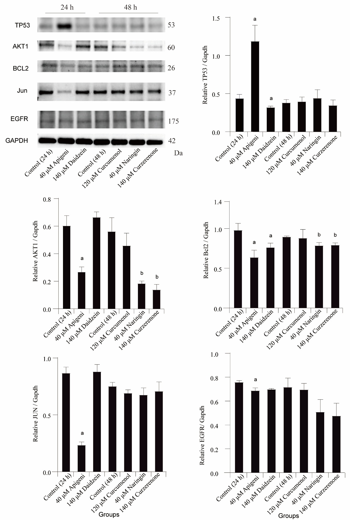
Figure 4 Apigenin is the main active monomer of JHD in promoting the apoptosis of HepG2 cells A: electrophoresis band images of AKT1, BCL2, TP53, JUN, EGFR, and GAPDH; B: bar chart of TP53 expression in each group; C: bar chart of AKT1 expression in each group; D: bar chart of BCL2 expression in each group; E: bar chart of JUN expression in each group; F: bar chart of EGFR expression in each group. The protein expressions of AKT1, BCL2, TP53, JUN, and EGFR were analyzed using Western blotting. Specifically, apigenin and daidzein were used to stimulate HepG2 cells in vitro for 24 h, while curcumenol, curzerenone, and naringin were applied to stimulate HepG2 cells in vitro for 48 h (n = 3). TP53: tumor protein 53; AKT1: serine-threonine kinase 1; BCL2: B-cell lymphoma 2; EGFR: epidermal growth factor receptor; JUN: Jun proto-oncogene; GAPDH: glyceraldehyde-3-phosphate dehydrogenase; HepG2: human hepatoma G2 cell line; JHD: Jianpi Huayu decoction. The statistical method used was independent samples t-tests. aP < 0.001, compared with the 24 h control group; bP < 0.05, compared with the 48 h control group.
Figure 4 Apigenin is the main active monomer of JHD in promoting the apoptosis of HepG2 cells A: electrophoresis band images of AKT1, BCL2, TP53, JUN, EGFR, and GAPDH; B: bar chart of TP53 expression in each group; C: bar chart of AKT1 expression in each group; D: bar chart of BCL2 expression in each group; E: bar chart of JUN expression in each group; F: bar chart of EGFR expression in each group. The protein expressions of AKT1, BCL2, TP53, JUN, and EGFR were analyzed using Western blotting. Specifically, apigenin and daidzein were used to stimulate HepG2 cells in vitro for 24 h, while curcumenol, curzerenone, and naringin were applied to stimulate HepG2 cells in vitro for 48 h (n = 3). TP53: tumor protein 53; AKT1: serine-threonine kinase 1; BCL2: B-cell lymphoma 2; EGFR: epidermal growth factor receptor; JUN: Jun proto-oncogene; GAPDH: glyceraldehyde-3-phosphate dehydrogenase; HepG2: human hepatoma G2 cell line; JHD: Jianpi Huayu decoction. The statistical method used was independent samples t-tests. aP < 0.001, compared with the 24 h control group; bP < 0.05, compared with the 48 h control group.
| [1] | Lau G, Obi S, Zhou J, et al. APASL clinical practice guidelines on systemic therapy for hepatocellular carcinoma-2024. Hepatol Int 2024; 18: 1661-83. |
| [2] | Llovet JM, Pinyol R, Yarchoan M, et al. Adjuvant and neoadjuvant immunotherapies in hepatocellular carcinoma. Nat Rev Clin Oncol 2024; 21: 294-311. |
| [3] | Li Y, Ji W, Wang C, et al. Poly l-Lactic acid nanofiber membrane effectively inhibits liver cancer cells growth and prevents postoperative residual cancer recurrence. ACS Appl Mater Interfaces 2025; 17: 689-700. |
| [4] | Eskander G, Abdelhamid SG, Wahdan SA, Radwan SM. Insights on the crosstalk among different cell death mechanisms. Cell Death Discov 2025; 11: 56. |
| [5] | Zhang MY, Zhang MY, Sun RQ, et al. Synergistic antitumor effect of oroxylin A and donafenib in hepatocellular carcinoma through tumor protein p53 signaling pathway activation. World J Gastroenterol 2026; 32: 113529. |
| [6] | Ai Y, Meng Y, Yan B, Zhou Q, Wang X. The biochemical pathways of apoptotic, necroptotic, pyroptotic, and ferroptotic cell death. Mol Cell 2024; 84: 170-9. |
| [7] | Jenča A, Mills DK, Ghasemi H, et al. Herbal therapies for cancer treatment: a review of phytotherapeutic efficacy. Biol Targets Ther 2024; 18: 229-55. |
| [8] | Shang Y, Zhao H. Research progress of Chinese medicinal monomers in the process of melanoma occurrence. Pharm Biol 2025; 63: 53-67. |
| [9] | Xie Y, Zhang Y, Wei X, et al. Jianpi Huayu decoction attenuates the immunosuppressive status of H22 hepatocellular carcinoma-bearing mice: by targeting myeloid-derived suppressor cells. Front Pharmacol 2020; 11: 16. |
| [10] | Chen JY, Chen SR, Luo XQ, et al. Effects of Jianpi Huayu decoction on Th1/Th2 immune balance in mice with liver cancer-related fatigue via the IL-27/STAT1 signaling pathway. Integr Cancer Ther 2024; 23: 15347354241263018. |
| [11] | Fang C, Liu S, Zhang S, et al. Jianpi Huayu decoction enhances the antitumor effect of doxorubicin via piezo1-mediated autophagy in hepatocellular carcinoma. Phytomedicine 2025; 143: 156908. |
| [12] | Huang Y, Zhou C, Wen H, et al. Jianpi-Huayu formula inhibits development of hepatocellular carcinoma by regulating expression of miR-602, which targets the RASSF1A gene. Integr Cancer Ther 2020; 19: 1534735419900804. |
| [13] | Qiu J, Zhang Y, Chen K, et al. Integrating serum pharmacochemistry, network pharmacology, metabolomics and 16S rRNA sequencing to explore the mechanism of total flavonoids from Flemingia philippinensis in treating collagen induced arthritis rats. Phytomedicine Int J Phytother Phytopharm 2025; 139: 156531. |
| [14] | Wang X, Yang X, Liu C, et al. Serum pharmacochemistry combined with network pharmacology reveals the hepatotoxicity mechanism of Alangium chinense (Lour.) Harms. J Ethnopharmacol 2025; 340: 119312. |
| [15] | Wang M, Yin F, Kong L, et al. Chinmedomics: a potent tool for the evaluation of Traditional Chinese Medicine efficacy and identification of its active components. Chin Med 2024; 19: 47. |
| [16] | Luo H, Wang M, Xu K, et al. Effect of Fushengong decoction on PTEN/PI3K/AKT/NF-κB pathway in rats with chronic renal failure via dual-dimension network pharmacology strategy. Front Pharmacol 2022; 13: 807651. |
| [17] | Liu Y, Yang X, Gan J, Chen S, Xiao ZX, Cao Y. CB-dock2: improved protein-ligand blind docking by integrating cavity detection, docking and homologous template fitting. Nucleic Acids Res 2022; 50: W159-64. |
| [18] | Chen M, Zhang S, Huang X, et al. The protective effects and mechanism of myricetin in liver diseases. Mol Med Rep 2025; 31: 87. |
| [19] | Oršolić N, Jazvinšćak Jembrek M. Potential strategies for overcoming drug resistance pathways using propolis and its polyphenolic/flavonoid compounds in combination with chemotherapy and radiotherapy. Nutrients 2024; 16: 3741. |
| [20] | Wang C, Feng X, Li W, et al. Apigenin as an emerging hepatoprotective agent: current status and future perspectives. Front Pharmacol 2024; 15: 1508060. |
| [21] | Naponelli V, Piscazzi A, Mangieri D. Cellular and molecular mechanisms modulated by genistein in cancer. Int J Mol Sci 2025; 26: 1114. |
| [22] | Zhang W, Zhuang X, Wu C, et al. Apigenin inhibits tumor angiogenesis by hindering microvesicle biogenesis via ARHGEF1. Cancer Lett 2024; 596: 216961. |
| [23] | Peng C, Zhang X, Zhou N, et al. Apigenin inhibits lipid metabolism of hepatocellular carcinoma cells by targeting the histone demethylase KDM1A. Phytomedicine Int J Phytother Phytopharm 2024; 135: 156024. |
| [24] | Mabrouk Zayed MM, Sahyon HA, Hanafy NAN, El-Kemary MA. The effect of encapsulated apigenin nanoparticles on HepG-2 cells through regulation of P53. Pharmaceutics 2022; 14: 1160. |
| [25] | Singh D, Khan MA, Mishra D, et al. Apigenin enhances sorafenib anti-tumour efficacy in hepatocellular carcinoma. Transl Oncol 2024; 43: 101920. |
| [26] | Korga A, Ostrowska M, Jozefczyk A, et al. Apigenin and hesperidin augment the toxic effect of doxorubicin against HepG2 cells. BMC Pharmacol Toxicol 2019; 20: 22. |
| [27] | Kan H, Wang P, Yang Y, et al. Apigenin inhibits proliferation and differentiation of cardiac fibroblasts through AKT/GSK3β signaling pathway. J Ethnopharmacol 2024; 334: 118518. |
| [28] | Yang J, Pi C, Wang G. Inhibition of PI3K/Akt/mTOR pathway by apigenin induces apoptosis and autophagy in hepatocellular carcinoma cells. Biomed Pharmacother Biomedecine Pharmacother 2018; 103: 699-707. |
| [29] | Chang JH, Cheng CW, Yang YC, et al. Downregulating CD26/DPPIV by apigenin modulates the interplay between Akt and Snail/Slug signaling to restrain metastasis of lung cancer with multiple EGFR statuses. J Exp Clin Cancer Res CR 2018; 37: 199. |
| [30] | Masuelli L, Marzocchella L, Quaranta A, et al. Apigenin induces apoptosis and impairs head and neck carcinomas EGFR/ErbB2 signaling. Front Biosci Landmark Ed 2011; 16: 1060-8. |
| [31] | Fu J, Zeng W, Chen M, et al. Apigenin suppresses tumor angiogenesis and growth via inhibiting HIF-1α expression in non-small cell lung carcinoma. Chem Biol Interact 2022; 361: 109966. |
| [32] | Zeng J, Xie H, Zhang ZL, et al. Apigenin regulates the migration, invasion, and autophagy of hepatocellular carcinoma cells by downregulating YAP. Neoplasma 2022; 69: 292-302. |
| [33] | Wang SM, Yang PW, Feng XJ, et al. Apigenin inhibits the growth of hepatocellular carcinoma cells by affecting the expression of microRNA transcriptome. Front Oncol 2021; 11: 657665. |
| [34] | Ganguly S, Dewanjee S, Sen R, et al. Apigenin-loaded galactose tailored PLGA nanoparticles: a possible strategy for liver targeting to treat hepatocellular carcinoma. Colloids Surf B Biointerfaces 2021; 204: 111778. |
| [35] | Khan F, Alam MW, Ramniwas S, Rautela I, Lakhanpal S, Pandey P. An updated review deciphering apigenin nanostructures as promising therapeutic efficiency in human carcinomas. Curr Med Chem 2025; 32: 2349-2367. |
| [36] | Cicek M, Unsal V, Emre A, Doganer A. Investigation of the effects of apigenin, a possible therapeutic agent, on cytotoxic and SWH pathway in colorectal cancer (HT29) cells. Adv Pharm Bull 2023; 13: 188-95. |
| [37] | Banjerdpongchai R, Wudtiwai B, Khawon P. Induction of human hepatocellular carcinoma HepG2 cell apoptosis by Naringin. Asian Pac J Cancer Prev APJCP 2016; 17: 3289-94. |
| [38] | Elsawy H, Algefare AI, Alfwuaires M, et al. Naringin alleviates methotrexate-induced liver injury in male albino rats and enhances its antitumor efficacy in HepG 2 cells. Biosci Rep 2020; 40: BSR20193686. |
| [39] | Xu C, Huang X, Huang Y, et al. Naringin induces apoptosis of gastric carcinoma cells via blocking the PI3K/AKT pathway and activating pro-death autophagy. Mol Med Rep 2021; 24: 772. |
| [40] | Tang S, Hu J, Meng Q, et al. Daidzein induced apoptosis via down-regulation of Bcl-2/Bax and triggering of the mitochondrial pathway in BGC-823 cells. Cell Biochem Biophys 2013; 65: 197-202. |
| [41] | Liang YS, Qi WT, Guo W, Wang CL, Hu ZB, Li AK. Genistein and daidzein induce apoptosis of colon cancer cells by inhibiting the accumulation of lipid droplets. Food Nutr Res 2018; 62: 1384. |
| [42] | Yanagihara K, Numoto M, Tauchi H, et al. Genetic status of p53 and induction of apoptosis by radiation or isoflavones in human gastric carcinoma cell lines. Int J Oncol 1996; 9: 95-102. |
| [43] | Wang BF, Wang JS, Lu JF, Kao TH, Chen BH. Antiproliferation effect and mechanism of prostate cancer cell lines as affected by isoflavones from soybean cake. J Agric Food Chem 2009; 57: 2221-32. |
| [44] | Ye R, Goodarzi AA, Kurz EU, et al. The isoflavonoids genistein and quercetin activate different stress signaling pathways as shown by analysis of site-specific phosphorylation of ATM, p53 and histone H2AX. DNA Repair 2004; 3: 235-44. |
| [45] | Han BJ, Li W, Jiang GB, et al. Effects of daidzein in regards to cytotoxicity in vitro, apoptosis, reactive oxygen species level, cell cycle arrest and the expression of caspase and Bcl-2 family proteins. Oncol Rep 2015; 34: 1115-20. |
| [46] | Liang J, Chen M, Yan G, et al. Donafenib activates the p53 signaling pathway in hepatocellular carcinoma, induces ferroptosis, and enhances cell apoptosis. Clin Exp Med 2025; 25: 29. |
| [47] | Sun Y, Wang M, Ling J, Wu Q, Han G, Zhou J. Curzerenone inactivates the nuclear factor-kappa B signaling to suppress malignancy and immune evasion in cervical cancer by targeting CSNK2B. Hum Cell 2024; 38: 35. |
| [48] | Syed Abdul Rahman SN, Abdul Wahab N, Abd Malek SN. In vitro morphological assessment of apoptosis induced by antiproliferative constituents from the rhizomes of curcuma zedoaria. Evid-Based Complement Altern Med ECAM 2013; 2013: 257108. |
| [49] | Liu XY, Chang YL, Wang XH, et al. An integrated approach to uncover anti-tumor active materials of curcumae rhizoma-sparganii rhizoma based on spectrum-effect relationship, molecular docking, and ADME evaluation. J Ethnopharmacol 2021; 280: 114439. |
| [50] | Han H, Wang L, Liu Y, et al. Combination of curcuma zedoary and kelp inhibits growth and metastasis of liver cancer in vivo and in vitro via reducing endogenous H2S levels. Food Funct 2019; 10: 224-34. |
| [51] | Zhou Y, Moon JH, Kim JT, et al. Curcumol metabolized by rat liver S9 fraction and orally administered in mouse suppressed the proliferation of colon cancer in vitro and in vivo. Food Sci Biotechnol 2024; 33: 171-80. |
| Viewed | ||||||
|
Full text |
|
|||||
|
Abstract |
|
|||||