
Journal of Traditional Chinese Medicine ›› 2026, Vol. 46 ›› Issue (4): 779-787.DOI: 10.19852/j.cnki.jtcm.2026.04.001
• Original Articles • Next Articles
Paeonol’s protective effects on H9C2 cells against hypoxia/ reoxygenation injury through the hypoxia-inducible factor 1-alpha/B-cell lymphoma-2/adenovirus E1B 19 kDa interacting protein 3 signaling pathway
ZHAO Chengguo, YIN Meifang, ZHOU Menghan, QIN Shuzhi( )
)
College of Life Science ,Zhuhai College of Science and Technology Zhuhai 519041, China
-
Received:2025-08-19Accepted:2026-01-30Online:2026-08-15Published:2026-08-08 -
Contact:Prof. QIN Shuzhi, College of Life Science, Zhuhai College of Science and Technology, Zhuhai 519041, China. qsz@zcst.edu.cn, Telephone: +86-13631282998
-
Supported by:Characteristic Innovation Project of Ordinary Colleges and Universities in Guangdong: Exploring the Mechanism of Paeoniflorin Against Myocardial Ischemia-reperfusion Injury Based on the Phosphatase and Tensin Homolog-induced Putative Kinase 1/Parkin Signaling Pathway(2018KTSCX307);Research Capacity Enhancement Project for Key Construction Disciplines of Guangdong Province: Study on the Pharmacological Effects and Molecular Mechanisms of Traditional Chinese Medicine Against Atherosclerosis Based on the Theory of "Li Shi Hua Zhuo"(2025ZDJS115);Tri-level Talents Development Project of Zhuhai College of Science and Technology

Cite this article
ZHAO Chengguo, YIN Meifang, ZHOU Menghan, QIN Shuzhi. Paeonol’s protective effects on H9C2 cells against hypoxia/ reoxygenation injury through the hypoxia-inducible factor 1-alpha/B-cell lymphoma-2/adenovirus E1B 19 kDa interacting protein 3 signaling pathway[J]. Journal of Traditional Chinese Medicine, 2026, 46(4): 779-787.
share this article
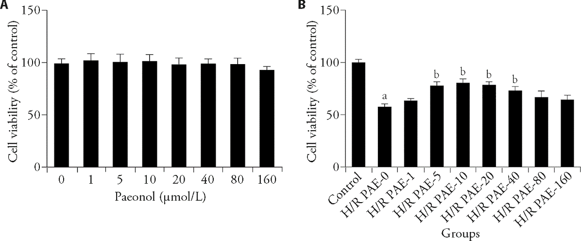
Figure 1 Impact of PAE on cell viability in H9C2 cells A: the cytotoxic effect of PAE at different concentrations (0, 1, 5, 10, 20, 40, 80, 160 μmol/L) on H9C2 cells was assessed by CCK8; B: effect of PAE on H/R-impaired cell viability was also tested by CCK8. Control group: cells were treated with vehicle; H/R + PAE: cells were treated with 12 h-hypoxia/12 h-reoxygenation and various concentrations (0, 1, 5, 10, 20, 40, 80, 160 μmol/L) of PAE. PAE: paeonol; CCK8: cell counting kit-8; H/R: hypoxia/reoxygenation. Statistical analyses were measured using unpaired t test (A) and one-way analysis of variance (B). Data are presented as mean ± standard error of the mean (n = 5). aP < 0.01, vs Control group; bP < 0.01, vs H/R group.
Figure 1 Impact of PAE on cell viability in H9C2 cells A: the cytotoxic effect of PAE at different concentrations (0, 1, 5, 10, 20, 40, 80, 160 μmol/L) on H9C2 cells was assessed by CCK8; B: effect of PAE on H/R-impaired cell viability was also tested by CCK8. Control group: cells were treated with vehicle; H/R + PAE: cells were treated with 12 h-hypoxia/12 h-reoxygenation and various concentrations (0, 1, 5, 10, 20, 40, 80, 160 μmol/L) of PAE. PAE: paeonol; CCK8: cell counting kit-8; H/R: hypoxia/reoxygenation. Statistical analyses were measured using unpaired t test (A) and one-way analysis of variance (B). Data are presented as mean ± standard error of the mean (n = 5). aP < 0.01, vs Control group; bP < 0.01, vs H/R group.
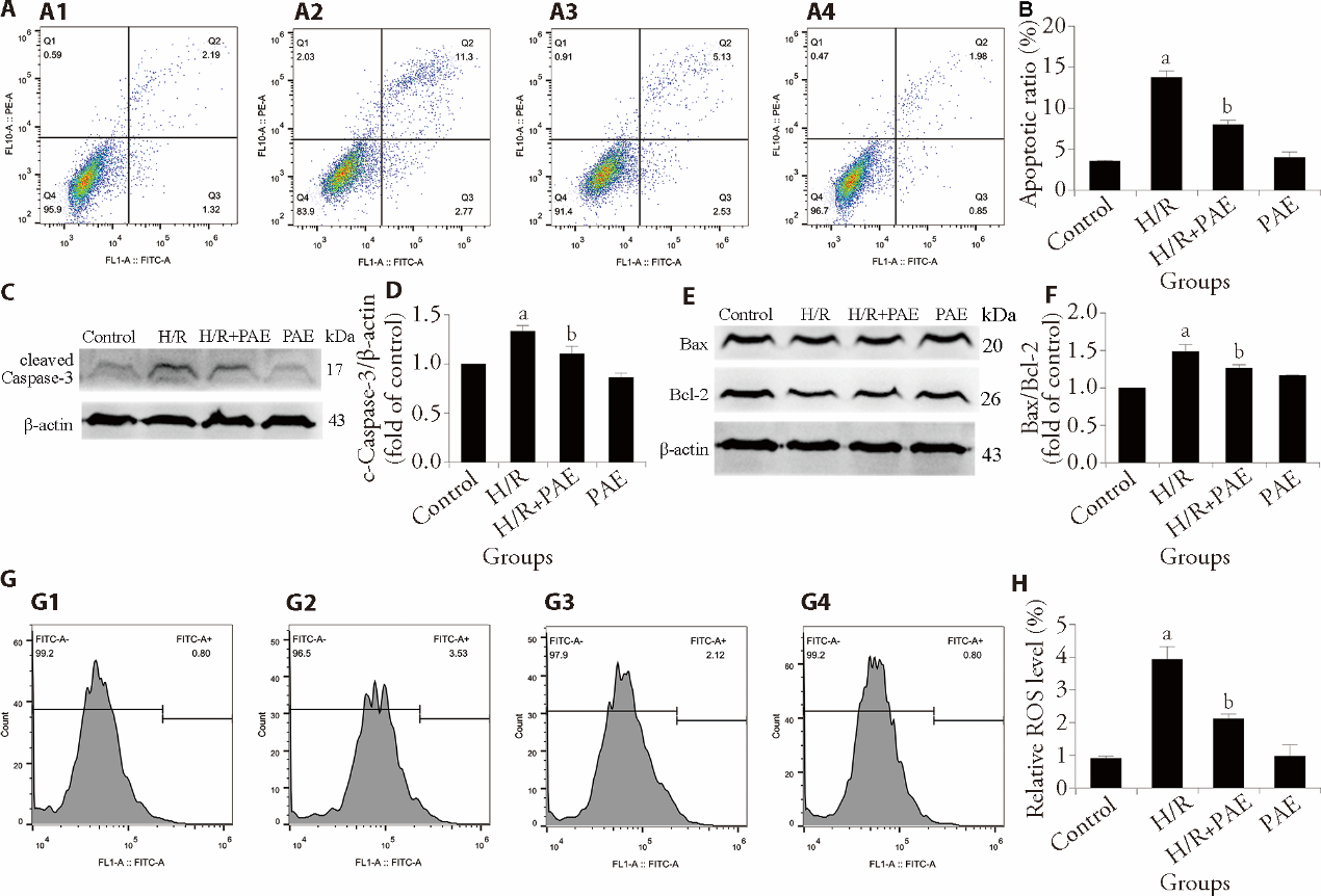
Figure 2 Effect of PAE on apoptosis in H/R-injured H9C2 cells A: the apoptosis rate of H9C2 cells was measured via flow cytometry. B: apoptosis rate of quantitative analysis; C: expression of cleaved caspase-3 was surveyed via Western blot; D: cleaved caspase-3 level of quantitative analysis; E: expression of Bax/Bcl-2 ratio was surveyed via Western blot; F: the Bax/Bcl-2 ratio of quantitative analysis; G: the ROS content in H9C2 cells was detected via flow cytometry; A1, G1: Control group; A2, G2: H/R group; A3, G3: H/R + PAE group; A4, G4: PAE group; H: ROS content of quantitative analysis. Control group: cells were treated with vehicle; H/R group: cells were treated with 12 h-hypoxia/12 h-reoxygenation; H/R+PAE group: cells were treated with 12 h-hypoxia/12 h-reoxygenation and 10 μmol/L Paeonol; PAE group: cells were treated with 10 μmol/L Paeonol. PAE: paeonol; H/R: hypoxia/reoxygenation; Bax: B-cell lymphoma 2-associated X protein; Bcl-2: B-cell lymphoma 2; ROS: reactive oxygen species. Statistical analyses were measured using one-way analysis of variance. Data are presented as mean ± standard error of the mean (n = 3). aP < 0.01 vs Control group; bP < 0.01 vs H/R group.
Figure 2 Effect of PAE on apoptosis in H/R-injured H9C2 cells A: the apoptosis rate of H9C2 cells was measured via flow cytometry. B: apoptosis rate of quantitative analysis; C: expression of cleaved caspase-3 was surveyed via Western blot; D: cleaved caspase-3 level of quantitative analysis; E: expression of Bax/Bcl-2 ratio was surveyed via Western blot; F: the Bax/Bcl-2 ratio of quantitative analysis; G: the ROS content in H9C2 cells was detected via flow cytometry; A1, G1: Control group; A2, G2: H/R group; A3, G3: H/R + PAE group; A4, G4: PAE group; H: ROS content of quantitative analysis. Control group: cells were treated with vehicle; H/R group: cells were treated with 12 h-hypoxia/12 h-reoxygenation; H/R+PAE group: cells were treated with 12 h-hypoxia/12 h-reoxygenation and 10 μmol/L Paeonol; PAE group: cells were treated with 10 μmol/L Paeonol. PAE: paeonol; H/R: hypoxia/reoxygenation; Bax: B-cell lymphoma 2-associated X protein; Bcl-2: B-cell lymphoma 2; ROS: reactive oxygen species. Statistical analyses were measured using one-way analysis of variance. Data are presented as mean ± standard error of the mean (n = 3). aP < 0.01 vs Control group; bP < 0.01 vs H/R group.
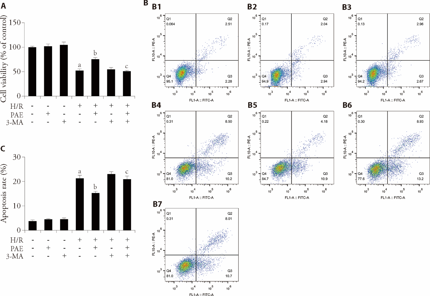
Figure 3 Effect of PAE and 3-MA on the autophagy flux and apoptosis induced by H/R injury in H9C2 cells A: the viability of PAE and 3-MA on H9C2 cells was measured by CCK8; B: the apoptosis rate of PAE and 3-MA on H9C2 cells was tested by flow cytometry; B1: Control group; B2: PAE group; B3: 3-MA group; B4: H/R group; B5: H/R + PAE group; B6: H/R + 3-MA group; B7: H/R + PAE + 3-MA group; C: apoptosis rate of quantitative analysis. Control group: cells were treated with vehicle; PAE group: cells were treated with 10 μmol/L Paeonol; 3-MA group: cells were treated with 5 mmol/L 3-Methyladenine; H/R group: cells were treated with 12 h-hypoxia/12 h-reoxygenation; H/R + PAE group: cells were treated with 12 h-hypoxia/12 h-reoxygenation and 10 μmol/L Paeonol; H/R + 3-MA group: cells were treated with 12 h-hypoxia/12 h-reoxygenation and 5 mmol/L 3-Methyladenine; H/R + PAE + 3-MA group: cells were treated with 12 h-hypoxia/12 h-reoxygenation, 10 μmol/L Paeonol and 5 mmol/L 3-Methyladenine. PAE: paeonol; H/R: hypoxia/reoxygenation; CCK8: cell counting kit-8; 3-MA: 3-methyladenine. Statistical analyses were measured using one-way analysis of variance. Data are presented as mean ± standard error of the mean (n = 3). aP < 0.01, vs Control group; bP <0.01, vs H/R group; cP < 0.01, vs H/R + PAE group.
Figure 3 Effect of PAE and 3-MA on the autophagy flux and apoptosis induced by H/R injury in H9C2 cells A: the viability of PAE and 3-MA on H9C2 cells was measured by CCK8; B: the apoptosis rate of PAE and 3-MA on H9C2 cells was tested by flow cytometry; B1: Control group; B2: PAE group; B3: 3-MA group; B4: H/R group; B5: H/R + PAE group; B6: H/R + 3-MA group; B7: H/R + PAE + 3-MA group; C: apoptosis rate of quantitative analysis. Control group: cells were treated with vehicle; PAE group: cells were treated with 10 μmol/L Paeonol; 3-MA group: cells were treated with 5 mmol/L 3-Methyladenine; H/R group: cells were treated with 12 h-hypoxia/12 h-reoxygenation; H/R + PAE group: cells were treated with 12 h-hypoxia/12 h-reoxygenation and 10 μmol/L Paeonol; H/R + 3-MA group: cells were treated with 12 h-hypoxia/12 h-reoxygenation and 5 mmol/L 3-Methyladenine; H/R + PAE + 3-MA group: cells were treated with 12 h-hypoxia/12 h-reoxygenation, 10 μmol/L Paeonol and 5 mmol/L 3-Methyladenine. PAE: paeonol; H/R: hypoxia/reoxygenation; CCK8: cell counting kit-8; 3-MA: 3-methyladenine. Statistical analyses were measured using one-way analysis of variance. Data are presented as mean ± standard error of the mean (n = 3). aP < 0.01, vs Control group; bP <0.01, vs H/R group; cP < 0.01, vs H/R + PAE group.
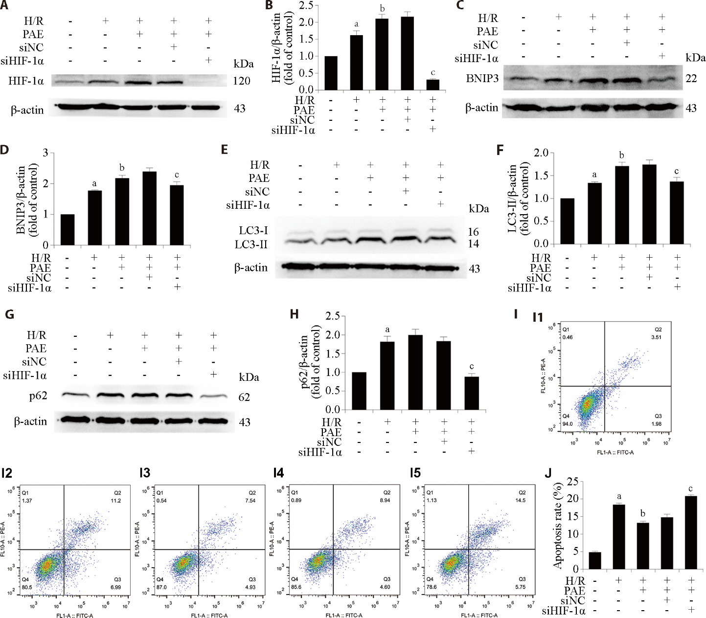
Figure 4 Effect of PAE and siHIF-1α interference on H/R-induced autophagy flux and apoptosis in H9C2 cells A: expression of the HIF-1α was evaluated by Western blot; B: HIF-1α expression of quantitative analysis; C: expression of the BNIP3 was evaluated by Western blot; D: BNIP3 expression of quantitative analysis; E: expression of the LC3 was evaluated by Western blot; F: LC3 expression of quantitative analysis; G: expression of the p62 was evaluated by Western blot; H: p62 expression of quantitative analysis; I: the apoptosis rate of H9C2 cells was measured by flow cytometry; I1: Control group; I2: H/R group; I3: H/R + PAE; I4: H/R + siNC + PAE; I5: H/R + siHIF-1α + PAE; J: apoptosis rate of quantitative analysis. Control group: cells were treated with vehicle; H/R group: cells were treated with 12 h-hypoxia/12 h-reoxygenation; H/R + PAE group: cells were treated with 12 h-hypoxia/12 h-reoxygenation and 10 μmol/L Paeonol; H/R + siNC + PAE: after siNC transfection, cells were treated with 10 μmol/L Paeonol; H/R + siHIF-1α + PAE: after siHIF-1α transfection, cells were treated with 10 μmol/L Paeonol. PAE: paeonol; H/R: hypoxia/reoxygenation; HIF-1α: hypoxia-inducible factor 1-alpha; BNIP3: B-cell lymphoma-2/adenovirus E1B 19 kDa interacting protein 3; LC3: microtubule-associated protein 1 light chain 3; p62: sequestosome-1; siNC: small interfering RNA negative control. Statistical analyses were measured using one-way analysis of variance. Data are presented as mean ± standard error of the mean (n = 3). aP < 0.01 vs Control group; bP < 0.01 vs H/R group; cP < 0.01 vs H/R + siNC + PAE group.
Figure 4 Effect of PAE and siHIF-1α interference on H/R-induced autophagy flux and apoptosis in H9C2 cells A: expression of the HIF-1α was evaluated by Western blot; B: HIF-1α expression of quantitative analysis; C: expression of the BNIP3 was evaluated by Western blot; D: BNIP3 expression of quantitative analysis; E: expression of the LC3 was evaluated by Western blot; F: LC3 expression of quantitative analysis; G: expression of the p62 was evaluated by Western blot; H: p62 expression of quantitative analysis; I: the apoptosis rate of H9C2 cells was measured by flow cytometry; I1: Control group; I2: H/R group; I3: H/R + PAE; I4: H/R + siNC + PAE; I5: H/R + siHIF-1α + PAE; J: apoptosis rate of quantitative analysis. Control group: cells were treated with vehicle; H/R group: cells were treated with 12 h-hypoxia/12 h-reoxygenation; H/R + PAE group: cells were treated with 12 h-hypoxia/12 h-reoxygenation and 10 μmol/L Paeonol; H/R + siNC + PAE: after siNC transfection, cells were treated with 10 μmol/L Paeonol; H/R + siHIF-1α + PAE: after siHIF-1α transfection, cells were treated with 10 μmol/L Paeonol. PAE: paeonol; H/R: hypoxia/reoxygenation; HIF-1α: hypoxia-inducible factor 1-alpha; BNIP3: B-cell lymphoma-2/adenovirus E1B 19 kDa interacting protein 3; LC3: microtubule-associated protein 1 light chain 3; p62: sequestosome-1; siNC: small interfering RNA negative control. Statistical analyses were measured using one-way analysis of variance. Data are presented as mean ± standard error of the mean (n = 3). aP < 0.01 vs Control group; bP < 0.01 vs H/R group; cP < 0.01 vs H/R + siNC + PAE group.
| 1. |
Vaduganathan M, Mensah GA, Turco JV, Fuster V, Roth GA. The global burden of cardiovascular diseases and risk: a compass for future health. J Am Coll Cardiol 2022; 80: 2361-71.
DOI PMID |
| 2. | Zhu M, Li Y, Xu Q, Wang W, Liu Y, Liu Y. Acute myocardial infarction: molecular pathogenesis, diagnosis, and clinical management. Med Comm (2020) 2025; 6: e70418. |
| 3. |
Fede MS, Daziani G, Tavoletta F, et al. Myocardial ischemia/reperfusion injury: molecular insights, forensic perspectives, and therapeutic horizons. Cells 2025; 14: 1509.
DOI URL |
| 4. |
Juricic S, Klac J, Stojkovic S, et al. Molecular and cellular mechanisms of myocardial ischemia and reperfusion injury: a narrative review. Cells 2026; 15: 265.
DOI URL |
| 5. |
He J, Liu D, Zhao L, et al. Myocardial ischemia/reperfusion injury: mechanisms of injury and implications for management (review). Exp Ther Med 2022; 23: 430.
DOI PMID |
| 6. |
Zhu Y, Liu F, Jian F, Rong Y. Recent progresses in the late stages of autophagy. Cell Insight 2024; 3: 100152.
DOI URL |
| 7. |
Lin PW, Chu ML, Liu HS. Autophagy and metabolism. Kaohsiung J Med Sci 2021; 37: 12-9.
DOI URL |
| 8. |
Zhou G, Zhang L, Bai W, et al. Devil or angel: a review of autophagy in ischemia-reperfusion injury. Medicine (Baltimore) 2025; 104: e45330.
DOI URL |
| 9. |
Shen L, Zhu Y, Chen Z, Shen F, Yu W, Zhang L. Isoliquiritigenin attenuates myocardial ischemia reperfusion through autophagy activation mediated by AMPK/mTOR/ULK1 signaling. BMC Cardiovasc Disord 2024; 24: 415.
DOI |
| 10. |
Xie M, Cho GW, Kong Y, et al. Activation of autophagic flux blunts cardiac ischemia/reperfusion injury. Circ Res 2021; 129: 435-50.
DOI PMID |
| 11. |
Liu XW, Lu MK, Zhong HT, Wang LH, Fu YP. Panax notoginseng saponins attenuate myocardial ischemia-reperfusion injury through the HIF-1α/BNIP3 pathway of autophagy. J Cardiovasc Pharmacol 2019; 73: 92-9.
DOI URL |
| 12. |
Zhu N, Li J, Li Y, et al. Berberine protects against simulated ischemia/reperfusion injury-induced H9C2 cardiomyocytes apoptosis in vitro and myocardial ischemia/reperfusion-induced apoptosis in vivo by regulating the mitophagy-mediated HIF-1α/BNIP3 pathway. Front Pharmacol 2020; 11: 367.
DOI PMID |
| 13. |
Li S, Jiang J, Fang J, et al. Naringin protects H9C 2 cardiomyocytes from chemical hypoxia-induced injury by promoting the autophagic flux via the activation of the HIF-1α/BNIP3 signaling pathway. Int J Mol Med 2021; 47: 102.
DOI URL |
| 14. |
Vellasamy S, Murugan D, Abas R, Alias A, Seng WY, Woon CK. Biological activities of paeonol in cardiovascular diseases: a review. Molecules 2021; 26: 4976..
DOI URL |
| 15. |
Ma X, Zhao X, Yang Y, et al. Paeonol inhibits NETs-mediated foam cell inflammation through the CitH3/NLRP3/caspase-1 signaling pathway in atherosclerosis. Int Immunopharmacol 2025; 151: 114340.
DOI URL |
| 16. |
Wang Y, Li BS, Zhang ZH, et al. Paeonol repurposing for cancer therapy: from mechanism to clinical translation. Biomed Pharmacother 2023; 165: 115277.
DOI URL |
| 17. |
Zheng J, Mao Z, Zhang J, Jiang L, Wang N. Paeonol pretreatment attenuates anoxia-reoxygenation induced injury in cardiac myocytes via a BRCA1 dependent pathway. Chem Pharm Bull (Tokyo) 2020; 68: 1163-9.
DOI URL |
| 18. |
Tsai CF, Su HH, Chen KM, et al. Paeonol protects against myocardial ischemia/reperfusion-induced injury by mediating apoptosis and autophagy crosstalk. Front Pharmacol 2021; 11: 586498.
DOI URL |
| 19. |
Qi C, Zhang J, Chen X, et al. Hypoxia stimulates neural stem cell proliferation by increasing HIF-1α expression and activating Wnt/β-catenin signaling. Cell Mol Biol (Noisy-le-grand) 2017; 63: 12-9.
DOI PMID |
| 20. |
Unnisa A, Greig NH, Kamal MA. Inhibition of caspase 3 and caspase 9 mediated apoptosis: a multimodal therapeutic target in traumatic brain injury. Curr Neuropharmacol 2023; 21: 1001-12.
DOI URL |
| 21. |
Li Z, Xiao G, Wang H, He S, Zhu Y. A preparation of Ginkgo biloba L. leaves extract inhibits the apoptosis of hippocampal neurons in post-stroke mice via regulating the expression of Bax/Bcl-2 and Caspase-3. J Ethnopharmacol 2021; 280: 114481.
DOI URL |
| 22. |
Bugger H, Pfeil K. Mitochondrial ROS in myocardial ischemia reperfusion and remodeling. Biochim Biophys Acta Mol Basis Dis 2020; 1866: 165768.
DOI URL |
| 23. |
Zhang Y, Liu D, Hu H, Zhang P, Xie R, Cui W. HIF-1α/BNIP3 signaling pathway-induced-autophagy plays protective role during myocardial ischemia-reperfusion injury. Biomed Pharmacother 2019; 120: 109464.
DOI URL |
| 24. |
Zhang S, Yan F, Luan F, et al. The pathological mechanisms and potential therapeutic drugs for myocardial ischemia reperfusion injury. Phytomedicine 2024; 129: 155649.
DOI URL |
| 25. |
Xiang Q, Yi X, Zhu XH, Wei X, Jiang DS. Regulated cell death in myocardial ischemia-reperfusion injury. Trends Endocrinol Metab 2024; 35: 219-34.
DOI URL |
| 26. |
Heusch G. Myocardial ischaemia-reperfusion injury and cardioprotection in perspective. Nat Rev Cardiol 2020; 17: 773-89.
DOI |
| 27. |
Zhang H, Hu H, Zhai C, Jing L, Tian H. Cardioprotective strategies after ischemia-reperfusion injury. Am J Cardiovasc Drugs 2024; 24: 5-18.
DOI |
| 28. |
Liu Y, Wu Z, Jin X, et al. Paeonol improves cardiac remodelling in MI mice by suppressing NOX2 mRNA expression to mitigate oxidative stress and mitochondrial dysfunction. J Cell Mol Med 2025; 29: e70563.
DOI URL |
| 29. |
Wu M, Yu Z, Li X, et al. Paeonol for the treatment of atherosclerotic cardiovascular disease: a pharmacological and mechanistic overview. Front Cardiovasc Med 2021; 8: 690116.
DOI URL |
| 30. |
Li X, Hu H, Yin J, Cheng W, Shi Y, Wang Y. Paeonol can improve hypoxic-induced H9c2 cells injury and ion channel activity by up-regulating the expression of CKIP-1. Tissue Cell 2024; 88: 102371.
DOI URL |
| 31. |
Korshunova AY, Blagonravov ML, Neborak EV, et al. BCL2-regulated apoptotic process in myocardial ischemia-reperfusion injury (Review). Int J Mol Med 2021; 47: 23-36.
DOI PMID |
| 32. |
McComb S, Chan PK, Guinot A, et al. Efficient apoptosis requires feedback amplification of upstream apoptotic signals by effector caspase-3 or -7. Sci Adv 2019; 5: eaau9433.
DOI URL |
| 33. |
Gong G, Wan W, Zhang X, Chen X, Yin J. Management of ROS and regulatory cell death in myocardial ischemia-reperfusion injury. Mol Biotechnol 2025; 67: 1765-83.
DOI |
| 34. |
Mokhtari B, Badalzadeh R. The potentials of distinct functions of autophagy to be targeted for attenuation of myocardial ischemia/reperfusion injury in preclinical studies: an up-to-date review. J Physiol Biochem 2021; 77: 377-404.
DOI PMID |
| 35. |
Xu H, Cheng J, He F. Cordycepin alleviates myocardial ischemia/reperfusion injury by enhancing autophagy via AMPK-mTOR pathway. J Physiol Biochem 2022; 78: 401-13.
DOI |
| 36. | Qu X, Chen X, Shi Q, Wang X, Wang D, Yang L. Resveratrol alleviates ischemia/reperfusion injury of diabetic myocardium via inducing autophagy. Exp Ther Med 2019; 18: 2719-25. |
| 37. |
Chen L, Lv Y, Wu H, et al. Gastrodin exerts perioperative myocardial protection by improving mitophagy through the PINK1/Parkin pathway to reduce myocardial ischemia-reperfusion injury. Phytomedicine 2024; 133: 155900.
DOI URL |
| 38. |
Wu Y, Zhang H, Wang Y, Zhang Y, Hong Z, Wang D. Sephin 1 enhances integrated stress response and autophagy to alleviate myocardial ischemia-reperfusion injury in mice. Biomed Pharmacother 2024; 176: 116869.
DOI URL |
| 39. | He H, Liu P, Li P. Dexmedetomidine ameliorates cardiac ischemia/reperfusion injury by enhancing autophagy through activation of the AMPK/SIRT3 pathway. Drug Des Devel Ther 2023; 17: 3205-18. |
| 40. |
Aljakna Khan A, Sabatasso S. Autophagy in myocardial ischemia and ischemia/reperfusion. Cardiovasc Pathol 2025; 74: 107691.
DOI URL |
| 41. |
Shang JN, Yu CG, Li R, et al. The nonautophagic functions of autophagy-related proteins. Autophagy 2024; 20: 720-34.
DOI URL |
| 42. |
Alcober-Boquet L, Zang T, Pietsch L, et al. The PB1 and the ZZ domain of the autophagy receptor p62/SQSTM1 regulate the interaction of p62/SQSTM1 with the autophagosome protein LC3B. Protein Sci 2024; 33: e4840.
DOI PMID |
| 43. |
Liu T, Zhang J, Li K, Deng L, Wang H. Combination of an autophagy inducer and an autophagy inhibitor: a smarter strategy emerging in cancer therapy. Front Pharmacol 2020; 11: 408.
DOI PMID |
| 44. | Youle R, Narendra D. Mechanisms of mitophagy. Nat Rev Mol Cell Biol 2011; 12: 9-14. |
| 45. |
Sousa Fialho MDL, Abd Jamil AH, Stannard GA, Heather LC. Hypoxia-inducible factor 1 signalling, metabolism and its therapeutic potential in cardiovascular disease. Biochim Biophys Acta Mol Basis Dis 2019; 1865: 831-43.
DOI URL |
| 46. |
Carmichael E, Reme AS, Bosco PJ, et al. Biological regulation of HIF-1α and its role in therapeutic angiogenesis for treatment of ischemic cardiovascular disease. Int J Mol Sci 2025; 26: 11236.
DOI URL |
| 47. |
Alanova P, Alan L, Opletalova B, et al. HIF-1α limits myocardial infarction by promoting mitophagy in mouse hearts adapted to chronic hypoxia. Acta Physiol (Oxf) 2024; 240: e14202.
DOI URL |
| 48. |
Gong N, Yang X, Li X, et al. MicroRNA-590-3p relieves hypoxia/reoxygenation induced cardiomyocytes apoptosis and autophagy by targeting HIF-1α. Exp Ther Med 2021; 22: 1077.
DOI PMID |
| 49. |
Fu ZJ, Wang ZY, Xu L, et al. HIF-1α-BNIP3-mediated mitophagy in tubular cells protects against renal ischemia/reperfusion injury. Redox Biol 2020; 36: 101671.
DOI URL |
| 50. |
Cheng S, Jian C, Yufang Z, et al. Protective effect of Dan Ze mixture against lipotoxic cardiomyopathy through activating B-cell lymphoma-2 adenovirus E1B 19 kDa-interacting protein 3/mitophagy signaling pathway. J Tradit Chin Med 2025; 45: 538-51.
DOI |
| 51. |
Zhang YN, Pang YX, Liu DW, Hu HJ, Xie RQ, Cui W. JMJD 5 attenuates oxygen-glucose deprivation and reperfusion-induced injury in cardiomyocytes through regulation of HIF-1α-BNIP3. Kaohsiung J Med Sci 2022; 38: 38-48.
DOI URL |
| No related articles found! |
| Viewed | ||||||
|
Full text |
|
|||||
|
Abstract |
|
|||||