
Journal of Traditional Chinese Medicine ›› 2026, Vol. 46 ›› Issue (4): 873-882.DOI: 10.19852/j.cnki.jtcm.2026.04.008
• Original Articles • Previous Articles Next Articles
Taohong Huazhuo decoction (桃红化浊汤) alleviates carbon tetrachloride-induced hepatic fibrosis in rats by inhibiting the nicotinamide adenine dinucleotide phosphate oxidase 4/nucleotide-binding oligomerization domain-like receptor family pyrin domain-containing 3 signaling pathway
REN Yuan1, CHEN Xiangni1, SHI Lei1, MAO Minghua1, LI Shufang1, YAO Jia2, HAO Jianmei1( )
)
- 1
Department of Hepatology ,Xi'an Hospital of Traditional Chinese Medicine Xi'an 710021, China
2Experimental Center ,Xi'an Hospital of Traditional Chinese Medicine Xi'an 710021, China
-
Received:2025-06-05Accepted:2025-11-04Online:2026-08-15Published:2026-08-08 -
Contact:Prof. HAO Jianmei, Department of Hepatology, Xi'an Hospital of Traditional Chinese Medicine, Xi'an 710021, China. hjmyzp@163.com,Telephone: +86-15129629909
-
Supported by:Youth Program of Xi'an Municipal Health Commission of China: Exploring the Molecular Mechanism of Taohong Huazhuo decoction in Improving Liver Fibrosis by Regulating the Nicotinamide Adenine Dinucleotide Phosphate Oxidase 2/Reactive Oxygen Species Signaling Pathway Based on the Theory of Damp-heat and Ministerial Fire(2024qn20);Cultivation Projects of the National Natural Science Foundation of Xi'an Traditional Chinese Medicine Hospital: Exploring the Molecular Mechanism of Taohong Huazhuo decoction Against Liver Fibrosis Based on Endoplasmic Reticulum Stress-mediated Ferroptosis(2024YJ02)

Cite this article
REN Yuan, CHEN Xiangni, SHI Lei, MAO Minghua, LI Shufang, YAO Jia, HAO Jianmei. Taohong Huazhuo decoction (桃红化浊汤) alleviates carbon tetrachloride-induced hepatic fibrosis in rats by inhibiting the nicotinamide adenine dinucleotide phosphate oxidase 4/nucleotide-binding oligomerization domain-like receptor family pyrin domain-containing 3 signaling pathway[J]. Journal of Traditional Chinese Medicine, 2026, 46(4): 873-882.
share this article
Table 1 THHZD improved liver function in rats with CCl4-induced hepatic fibrosis ($\bar{x}±s$)
| Group | n | ALT (U/L) | AST (U/L) | TBIL (μmol/L) |
|---|---|---|---|---|
| Normal | 8 | 52.23±7.17 | 156.00±28.65 | 0.70±0.14a |
| Model | 8 | 437.35±59.65a | 538.50±99.67a | 6.99±1.21 |
| Colchicine | 8 | 279.70±112.33b | 319.75±107.62c | 4.49±1.49b |
| THHZD-L | 8 | 309.38±110.19 | 371.00±98.33b | 5.01±1.50 |
| THHZD-M | 8 | 279.65±34.77b | 296.50±56.20c | 4.47±0.88b |
| THHZD-H | 8 | 233.05±49.9c | 307.50±86.34c | 3.45±1.75c |
Table 1 THHZD improved liver function in rats with CCl4-induced hepatic fibrosis ($\bar{x}±s$)
| Group | n | ALT (U/L) | AST (U/L) | TBIL (μmol/L) |
|---|---|---|---|---|
| Normal | 8 | 52.23±7.17 | 156.00±28.65 | 0.70±0.14a |
| Model | 8 | 437.35±59.65a | 538.50±99.67a | 6.99±1.21 |
| Colchicine | 8 | 279.70±112.33b | 319.75±107.62c | 4.49±1.49b |
| THHZD-L | 8 | 309.38±110.19 | 371.00±98.33b | 5.01±1.50 |
| THHZD-M | 8 | 279.65±34.77b | 296.50±56.20c | 4.47±0.88b |
| THHZD-H | 8 | 233.05±49.9c | 307.50±86.34c | 3.45±1.75c |

Figure 1 THHZD inhibited CCL4-induced hepatic fibrosis and hepatic stellate cell activation in rats A: hematoxylin and eosin staining of hepatic tissues (× 100, scale bars = 100 μm); B: Western blot detection of collagen I protein expression in hepatic tissues; C: densitometric quantification of collagen I in hepatic tissues; D: masson's trichrome staining of hepatic tissues (× 100, scale bars = 100 μm); E: interstitial collagen was determined using Masson's trichrome staining; F: images of immunohistochemistry staining of α-SMA (× 200, scale bars = 40 μm); G: statistical analysis of immunohistochemistry staining of α-SMA; A1, D1, F1: normal group; A2, D2, F2: model group; A3, D3, F3: colchicine group; A4, D4, F4: THHZD-L group; A5, D5, F5: THHZD-M group; A6, D6, F6: THHZD-H group. Normal group: daily saline gavage for 4 weeks; Model group: daily saline gavage for 4 weeks; Colchicine group: daily intragastric colchicine (0.1 mg/kg) for 4 weeks; THHZD-L group: daily intragastric THHZD (3.285 g/kg) for 4 weeks; THHZD-M group: daily intragastric THHZD (6.57 g/kg) for 4 weeks; THHZD-H group: daily intragastric THHZD (13.14 g/kg) for 4 weeks. α-SMA: alpha-smooth muscle actin; THHZD-L: low-dose of Taohong Huazhuo decoction; THHZD-M: medium-dose of Taohong Huazhuo decoction; THHZD-H: high-dose of Taohong Huazhuo decoction. Statistical comparisons among groups were conducted via one-way analysis of variance, followed by Tukey’s test for pairwise multiple comparisons. All data are expressed as mean ± standard deviation (n = 3). Compared to the normal group, aP < 0.01; compared to the model group, bP < 0.01 and cP < 0.05.
Figure 1 THHZD inhibited CCL4-induced hepatic fibrosis and hepatic stellate cell activation in rats A: hematoxylin and eosin staining of hepatic tissues (× 100, scale bars = 100 μm); B: Western blot detection of collagen I protein expression in hepatic tissues; C: densitometric quantification of collagen I in hepatic tissues; D: masson's trichrome staining of hepatic tissues (× 100, scale bars = 100 μm); E: interstitial collagen was determined using Masson's trichrome staining; F: images of immunohistochemistry staining of α-SMA (× 200, scale bars = 40 μm); G: statistical analysis of immunohistochemistry staining of α-SMA; A1, D1, F1: normal group; A2, D2, F2: model group; A3, D3, F3: colchicine group; A4, D4, F4: THHZD-L group; A5, D5, F5: THHZD-M group; A6, D6, F6: THHZD-H group. Normal group: daily saline gavage for 4 weeks; Model group: daily saline gavage for 4 weeks; Colchicine group: daily intragastric colchicine (0.1 mg/kg) for 4 weeks; THHZD-L group: daily intragastric THHZD (3.285 g/kg) for 4 weeks; THHZD-M group: daily intragastric THHZD (6.57 g/kg) for 4 weeks; THHZD-H group: daily intragastric THHZD (13.14 g/kg) for 4 weeks. α-SMA: alpha-smooth muscle actin; THHZD-L: low-dose of Taohong Huazhuo decoction; THHZD-M: medium-dose of Taohong Huazhuo decoction; THHZD-H: high-dose of Taohong Huazhuo decoction. Statistical comparisons among groups were conducted via one-way analysis of variance, followed by Tukey’s test for pairwise multiple comparisons. All data are expressed as mean ± standard deviation (n = 3). Compared to the normal group, aP < 0.01; compared to the model group, bP < 0.01 and cP < 0.05.

Figure 2 THHZD inhibits NOX4-mediated oxidative stress in the livers of rats with CCl4-induced hepatic fibrosis A: ROS expression in hepatic tissues was detected by immunofluorescence staining (× 200, scale bars = 50 μm). A1, A7, A13: normal group; A2, A8, A14: model group; A3, A9, A15: colchicine group; A4, A10, A16: THHZD-L group; A5, A11, A17: THHZD-M group; A6, A12, A18: THHZD-H group; B: statistical analysis of immunofluorescence of ROS; C: western blot detection of NOX4 protein expression in hepatic tissues; D: densitometric quantification of NOX4 in hepatic tissues; E: the levels of MDA in hepatic tissues were determined using ELISA kits; F: the levels of GSH in hepatic tissues were determined using ELISA kits. Normal group: daily saline gavage for 4 weeks; Model group: daily saline gavage for 4 weeks; Colchicine group: daily intragastric colchicine (0.1 mg/kg) for 4 weeks; THHZD-L group: daily intragastric THHZD (3.285 g/kg) for 4 weeks; THHZD-M group: daily intragastric THHZD (6.57 g/kg) for 4 weeks; THHZD-H group: daily intragastric THHZD (13.14 g/kg) for 4 weeks. GSH: glutathione; MDA: malondialdehyde; IL-1β: interleukin-1β; IL-18: interleukin-18; ROS: reactive oxygen species; NOX4: nicotinamide adenine dinucleotide phosphate oxidase 4; THHZD-L: low-dose of Taohong Huazhuo decoction; THHZD-M: medium-dose of Taohong Huazhuo decoction; THHZD-H: high-dose of Taohong Huazhuo decoction; ELISA: enzyme-linked immunosorbent assay. Statistical comparisons among groups were conducted via one-way analysis of variance, followed by Tukey’s test for pairwise multiple comparisons. All data are expressed as mean ± standard deviation (n = 3). Compared to the normal group, aP < 0.01; compared to the model group, bP < 0.01 and cP< 0.05.
Figure 2 THHZD inhibits NOX4-mediated oxidative stress in the livers of rats with CCl4-induced hepatic fibrosis A: ROS expression in hepatic tissues was detected by immunofluorescence staining (× 200, scale bars = 50 μm). A1, A7, A13: normal group; A2, A8, A14: model group; A3, A9, A15: colchicine group; A4, A10, A16: THHZD-L group; A5, A11, A17: THHZD-M group; A6, A12, A18: THHZD-H group; B: statistical analysis of immunofluorescence of ROS; C: western blot detection of NOX4 protein expression in hepatic tissues; D: densitometric quantification of NOX4 in hepatic tissues; E: the levels of MDA in hepatic tissues were determined using ELISA kits; F: the levels of GSH in hepatic tissues were determined using ELISA kits. Normal group: daily saline gavage for 4 weeks; Model group: daily saline gavage for 4 weeks; Colchicine group: daily intragastric colchicine (0.1 mg/kg) for 4 weeks; THHZD-L group: daily intragastric THHZD (3.285 g/kg) for 4 weeks; THHZD-M group: daily intragastric THHZD (6.57 g/kg) for 4 weeks; THHZD-H group: daily intragastric THHZD (13.14 g/kg) for 4 weeks. GSH: glutathione; MDA: malondialdehyde; IL-1β: interleukin-1β; IL-18: interleukin-18; ROS: reactive oxygen species; NOX4: nicotinamide adenine dinucleotide phosphate oxidase 4; THHZD-L: low-dose of Taohong Huazhuo decoction; THHZD-M: medium-dose of Taohong Huazhuo decoction; THHZD-H: high-dose of Taohong Huazhuo decoction; ELISA: enzyme-linked immunosorbent assay. Statistical comparisons among groups were conducted via one-way analysis of variance, followed by Tukey’s test for pairwise multiple comparisons. All data are expressed as mean ± standard deviation (n = 3). Compared to the normal group, aP < 0.01; compared to the model group, bP < 0.01 and cP< 0.05.
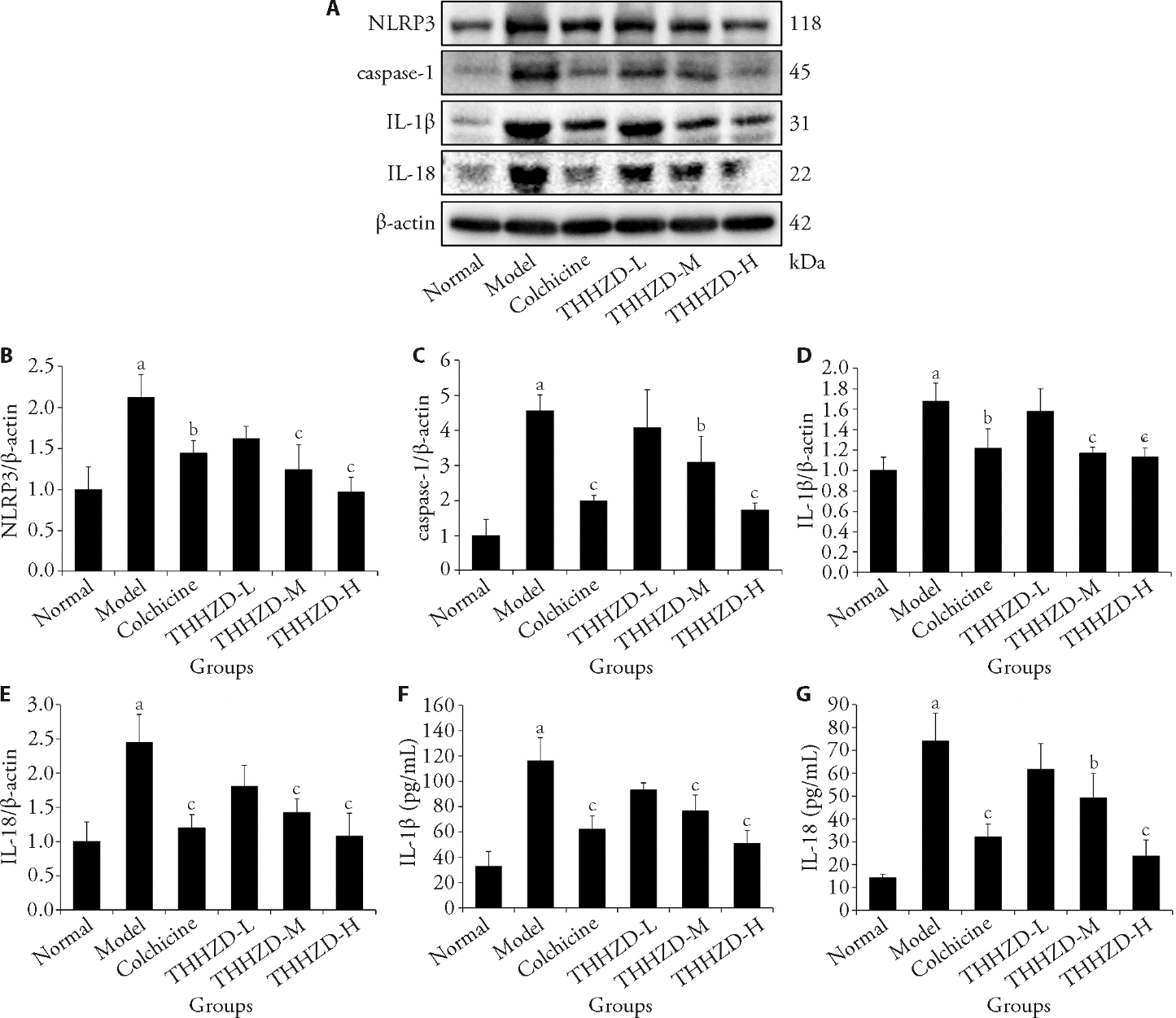
Figure 3 THHZD inhibits hepatic inflammasome activation in rats with CCl4-induced hepatic fibrosis A: Western blot detection of NLRP3, caspase-1, IL-1β, and IL-18 protein expression in hepatic tissues; B: densitometric quantification of NLRP3 in hepatic tissues; C: densitometric quantification of caspase-1 in hepatic tissues; D: densitometric quantification of IL-1β in hepatic tissues; E: densitometric quantification of IL-8 in hepatic tissues; F: the levels of serum IL-1β were determined using ELISA kits; G: the levels of serum IL-18 were determined using ELISA kits; Normal group: daily saline gavage for 4 weeks; Model group: daily saline gavage for 4 weeks; Colchicine group: daily intragastric colchicine (0.1 mg/kg) for 4 weeks; THHZD-L group: daily intragastric THHZD (3.285 g/kg) for 4 weeks; THHZD-M group: daily intragastric THHZD (6.57 g/kg) for 4 weeks; THHZD-H group: daily intragastric THHZD (13.14 g/kg) for 4 weeks. IL-1β: interleukin-1β; IL-18: interleukin-18; NLRP3: nucleotide-binding oligomerization domain-like receptor family pyrin domain-containing 3; THHZD-L: low-dose of Taohong Huazhuo decoction; THHZD-M: medium-dose of Taohong Huazhuo decoction; THHZD-H: high-dose of Taohong Huazhuo decoction; ELISA: enzyme-linked immunosorbent assay. Statistical comparisons among groups were conducted via one-way analysis of variance, followed by Tukey’s test for pairwise multiple comparisons. All data are expressed as mean ± standard deviation (n = 3). Compared to the normal group, aP < 0.01; compared to the model group, bP < 0.05 and cP< 0.01.
Figure 3 THHZD inhibits hepatic inflammasome activation in rats with CCl4-induced hepatic fibrosis A: Western blot detection of NLRP3, caspase-1, IL-1β, and IL-18 protein expression in hepatic tissues; B: densitometric quantification of NLRP3 in hepatic tissues; C: densitometric quantification of caspase-1 in hepatic tissues; D: densitometric quantification of IL-1β in hepatic tissues; E: densitometric quantification of IL-8 in hepatic tissues; F: the levels of serum IL-1β were determined using ELISA kits; G: the levels of serum IL-18 were determined using ELISA kits; Normal group: daily saline gavage for 4 weeks; Model group: daily saline gavage for 4 weeks; Colchicine group: daily intragastric colchicine (0.1 mg/kg) for 4 weeks; THHZD-L group: daily intragastric THHZD (3.285 g/kg) for 4 weeks; THHZD-M group: daily intragastric THHZD (6.57 g/kg) for 4 weeks; THHZD-H group: daily intragastric THHZD (13.14 g/kg) for 4 weeks. IL-1β: interleukin-1β; IL-18: interleukin-18; NLRP3: nucleotide-binding oligomerization domain-like receptor family pyrin domain-containing 3; THHZD-L: low-dose of Taohong Huazhuo decoction; THHZD-M: medium-dose of Taohong Huazhuo decoction; THHZD-H: high-dose of Taohong Huazhuo decoction; ELISA: enzyme-linked immunosorbent assay. Statistical comparisons among groups were conducted via one-way analysis of variance, followed by Tukey’s test for pairwise multiple comparisons. All data are expressed as mean ± standard deviation (n = 3). Compared to the normal group, aP < 0.01; compared to the model group, bP < 0.05 and cP< 0.01.
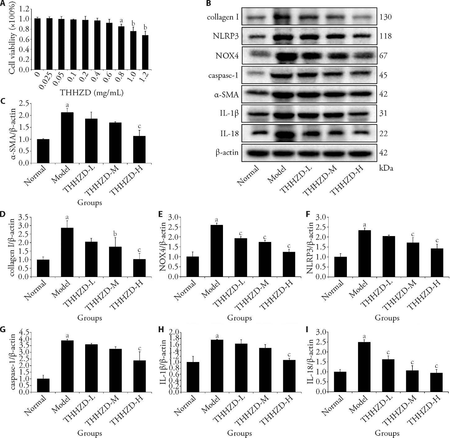
Figure 4 THHZD effectively suppresses the activation of HSCs via blocking the activation of the NOX4/NLRP3 signaling pathway in vitro A: effect of THHZD on the viability of HSC-T6 cells was evaluated using CCK-8 assay; B: Western blot detection of α-SMA, collagen I, NOX4, NLRP3, caspase-1, IL-1β, and IL-18 protein expression in HSC-T6 cells; C: densitometric quantification of α-SMA; D: densitometric quantification of collagen I; E: densitometric quantification of NOX4; F: densitometric quantification of NLRP3; G: densitometric quantification of caspase-1; H: densitometric quantification of IL-1β; I: densitometric quantification of IL-18; Normal group: no treatment; Model group: 10 ng/mL TGF-β1, 24 h; THHZD-L: 10 ng/mL TGF-β1 + 0.15 mg/mL THHZD, 24 h; THHZD-M: 10 ng/mL TGF-β1 + 0.3 mg/mL THHZD, 24 h; THHZD-H: 10 ng/mL TGF-β1 + 0.6 mg/mL THHZD, 24 h. IL-1β: interleukin-1β; IL-18: interleukin-18; NLRP3: nucleotide-binding oligomerization domain-like receptor family pyrin domain-containing 3; NOX4: nicotinamide adenine dinucleotide phosphate oxidase 4; α-SMA: alpha-smooth muscle actin; HSCs: hepatic stellate cells; TGF-β1: transforming growth factor-β1; CCK-8: cell counting kit-8; THHZD-L: low-dose of Taohong Huazhuo decoction; THHZD-M: medium-dose of Taohong Huazhuo decoction; THHZD-H: high-dose of Taohong Huazhuo decoction. Statistical comparisons among groups were conducted via one-way analysis of variance, followed by Tukey’s test for pairwise multiple comparisons. All data are expressed as mean ± standard deviation (n = 3). Compared to the normal group, aP < 0.01; compared to the model group, bP < 0.05 and cP < 0.01.
Figure 4 THHZD effectively suppresses the activation of HSCs via blocking the activation of the NOX4/NLRP3 signaling pathway in vitro A: effect of THHZD on the viability of HSC-T6 cells was evaluated using CCK-8 assay; B: Western blot detection of α-SMA, collagen I, NOX4, NLRP3, caspase-1, IL-1β, and IL-18 protein expression in HSC-T6 cells; C: densitometric quantification of α-SMA; D: densitometric quantification of collagen I; E: densitometric quantification of NOX4; F: densitometric quantification of NLRP3; G: densitometric quantification of caspase-1; H: densitometric quantification of IL-1β; I: densitometric quantification of IL-18; Normal group: no treatment; Model group: 10 ng/mL TGF-β1, 24 h; THHZD-L: 10 ng/mL TGF-β1 + 0.15 mg/mL THHZD, 24 h; THHZD-M: 10 ng/mL TGF-β1 + 0.3 mg/mL THHZD, 24 h; THHZD-H: 10 ng/mL TGF-β1 + 0.6 mg/mL THHZD, 24 h. IL-1β: interleukin-1β; IL-18: interleukin-18; NLRP3: nucleotide-binding oligomerization domain-like receptor family pyrin domain-containing 3; NOX4: nicotinamide adenine dinucleotide phosphate oxidase 4; α-SMA: alpha-smooth muscle actin; HSCs: hepatic stellate cells; TGF-β1: transforming growth factor-β1; CCK-8: cell counting kit-8; THHZD-L: low-dose of Taohong Huazhuo decoction; THHZD-M: medium-dose of Taohong Huazhuo decoction; THHZD-H: high-dose of Taohong Huazhuo decoction. Statistical comparisons among groups were conducted via one-way analysis of variance, followed by Tukey’s test for pairwise multiple comparisons. All data are expressed as mean ± standard deviation (n = 3). Compared to the normal group, aP < 0.01; compared to the model group, bP < 0.05 and cP < 0.01.
| 1. |
Li L, Zhu Z. Pharmacological modulation of ferroptosis as a therapeutic target for liver fibrosis. Front Pharmacol 2022; 13: 1071844.
DOI URL |
| 2. |
Lee YS, Seki E. In vivo and in vitro models to study liver fibrosis: mechanisms and limitations. Cell Mol Gastroenterol Hepatol 2023; 16: 355-67.
DOI URL |
| 3. | Berumen J, Baglieri J, Kisseleva T, Mekeel K. Liver fibrosis: pathophysiology and clinical implications. WIREs Mech Dis 2021; 13: e1499. |
| 4. |
Hijazi N, Rockey DC, Shi Z. The cellular microenvironment and cytoskeletal actin dynamics in liver fibrogenesis. Biocell 2022; 46: 2003-7.
DOI PMID |
| 5. |
Ali N, Ferrao K, Mehta KJ. Liver iron loading in alcohol-associated liver disease. Am J Pathol 2023; 193: 1427-39.
DOI URL |
| 6. |
Herranz-Itúrbide M, López-Luque J, Gonzalez-Sanchez E, et al. NADPH oxidase 4 (Nox4) deletion accelerates liver regeneration in mice. Redox Biol 2021; 40: 101841.
DOI URL |
| 7. | Matuz-Mares D, Vázquez-Meza H, Vilchis-Landeros MM. NOX as a therapeutic target in liver disease. Antioxidants (Basel) 2022; 11: 2038. |
| 8. | Zhai L, Pei H, Yang Y, Zhu Y, Ruan S. NOX4 promotes Kupffer cell inflammatory response via ROS-NLRP3 to aggravate liver inflammatory injury in acute liver injury. Aging (Albany NY) 2022; 14: 6905-16. |
| 9. |
Ramos-Tovar E, Muriel P. NLRP3 inflammasome in hepatic diseases: a pharmacological target. Biochem Pharmacol 2023; 217: 115861.
DOI URL |
| 10. |
Sun M, Zhang Y, Guo A, Xia Z, Peng L. Progress in the correlation between inflammasome NLRP3 and liver fibrosis. J Clin Transl Hepatol 2024; 12: 191-200.
DOI PMID |
| 11. |
Liu HL, Lü J, Zhao ZM, et al. Fuzhenghuayu decoction ameliorates hepatic fibrosis by attenuating experimental sinusoidal capillarization and liver angiogenesis. Sci Rep 2019; 9: 18719.
DOI |
| 12. |
Li Z, Zhu J, Ouyang H. Research progress of Traditional Chinese Medicine in improving hepatic fibrosis based on inhibiting pathological angiogenesis. Front Pharmacol 2023; 14: 1303012.
DOI URL |
| 13. | Chen XN, Li SF, Yuan C. Clinical efficacy and safety of Taohong Huazhuo decoction combined with entecavir in the treatment of chronic hepatitis B cirrhosis (dampness-heat accumulation syndrome). Zhong Guo Gan Zang Bing Za Zhi 2021; 13: 9-15. |
| 14. |
Bai Y, Liu J, Zhao J, et al. Changes and influencing factors of liver volume after transjugular intrahepatic portosystemic shunt: a retrospective cohort study. Quant Imaging Med Surg 2023; 13: 3029-39.
DOI URL |
| 15. |
Li X, Chen R, Kemper S, Brigstock DR. Production, exacerbating effect, and EV-mediated transcription of hepatic CCN2 in NASH: icmplications for diagnosis and therapy of NASH fibrosis. Int J Mol Sci 2023; 24: 12823.
DOI URL |
| 16. |
Liu X, Xu J, Rosenthal S, et al. Identification of lineage-specific transcription factors that prevent activation of hepatic stellate cells and promote fibrosis resolution. Gastroenterology 2020; 158: 1728-44.e14.
DOI PMID |
| 17. |
Nanda I, Schröder SK, Steinlein C, et al. Rat hepatic stellate cell line CFSC-2G: genetic markers and short tandem repeat profile useful for cell line authentication. Cells 2022; 11: 2900.
DOI URL |
| 18. | Xia XC, Xue SP, Song GY, et al. Anti-oxidative and immunological role of Cyclocarya paliurus polysaccharide on the liver injury of diabetic rats. J Tradit Chin Med 2024; 44: 1146-52. |
| 19. |
Zhang X, Zhang J, Zhou Z, et al. Integrated network pharmacology, metabolomics, and transcriptomics of Huanglian-Hongqu herb pair in non-alcoholic fatty liver disease. J Ethnopharmacol 2024; 325: 117828.
DOI URL |
| 20. |
Bao X, Li J, Ren C, et al. Aucubin ameliorates liver fibrosis and hepatic stellate cells activation in diabetic mice via inhibiting ER stress-mediated IRE1α/TXNIP/NLRP 3 inflammasome through NOX4/ROS pathway. Chem Biol Interact 2022; 365: 110074.
DOI URL |
| 21. |
Nie Y, Liu Q, Zhang W, Wan Y, Huang C, Zhu X. Ursolic acid reverses liver fibrosis by inhibiting NOX4/NLRP3 inflammasome pathways and bacterial dysbiosis. Gut Microbes 2021; 13: 1972746.
DOI URL |
| [1] | Jiang Hui, Gao Jiarong, Wang Yongzhong, Zhang Jiafu, Han Yanquan, Wei Liangbing, Liu Xiaochuang, Wu Jian. Panax notoginseng saponins ameliorates experimental hepatic fibrosis and hepatic stellate cell proliferation by inhibiting the Jak2/Stat3 pathways [J]. Journal of Traditional Chinese Medicine, 2016, 36(02): 217-224. |
| Viewed | ||||||
|
Full text |
|
|||||
|
Abstract |
|
|||||